Calcium »
PDB 7ucy-7uwv »
7ulk »
Calcium in PDB 7ulk: Human TRAP1 Nm in Complex with 42C
Protein crystallography data
The structure of Human TRAP1 Nm in Complex with 42C, PDB code: 7ulk
was solved by
T.R.Stachowski,
S.Nithianantham,
M.Vanarotti,
M.Fischer,
with X-Ray Crystallography technique. A brief refinement statistics is given in the table below:
| Resolution Low / High (Å) | 45.21 / 2.34 |
| Space group | P 1 21 1 |
| Cell size a, b, c (Å), α, β, γ (°) | 69.624, 89.399, 95.599, 90, 111.56, 90 |
| R / Rfree (%) | 22.3 / 26.8 |
Calcium Binding Sites:
The binding sites of Calcium atom in the Human TRAP1 Nm in Complex with 42C
(pdb code 7ulk). This binding sites where shown within
5.0 Angstroms radius around Calcium atom.
In total 2 binding sites of Calcium where determined in the Human TRAP1 Nm in Complex with 42C, PDB code: 7ulk:
Jump to Calcium binding site number: 1; 2;
In total 2 binding sites of Calcium where determined in the Human TRAP1 Nm in Complex with 42C, PDB code: 7ulk:
Jump to Calcium binding site number: 1; 2;
Calcium binding site 1 out of 2 in 7ulk
Go back to
Calcium binding site 1 out
of 2 in the Human TRAP1 Nm in Complex with 42C
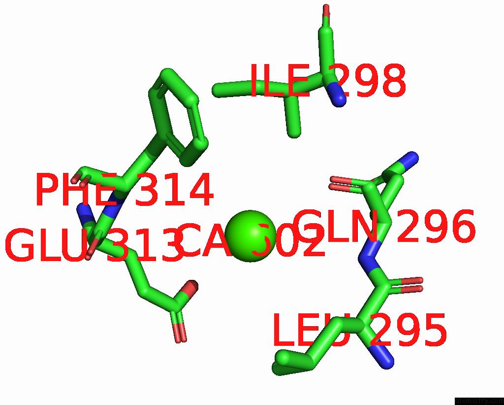
Mono view
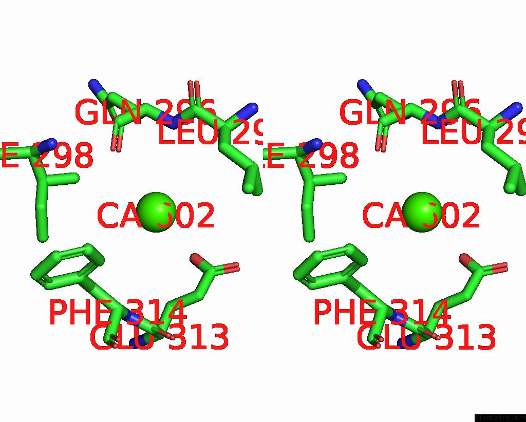
Stereo pair view
Mono view
Stereo pair view
A full contact list of Calcium with other atoms in the Ca binding
site number 1 of Human TRAP1 Nm in Complex with 42C within 5.0Å range:
|
Calcium binding site 2 out of 2 in 7ulk
Go back to
Calcium binding site 2 out
of 2 in the Human TRAP1 Nm in Complex with 42C

Mono view
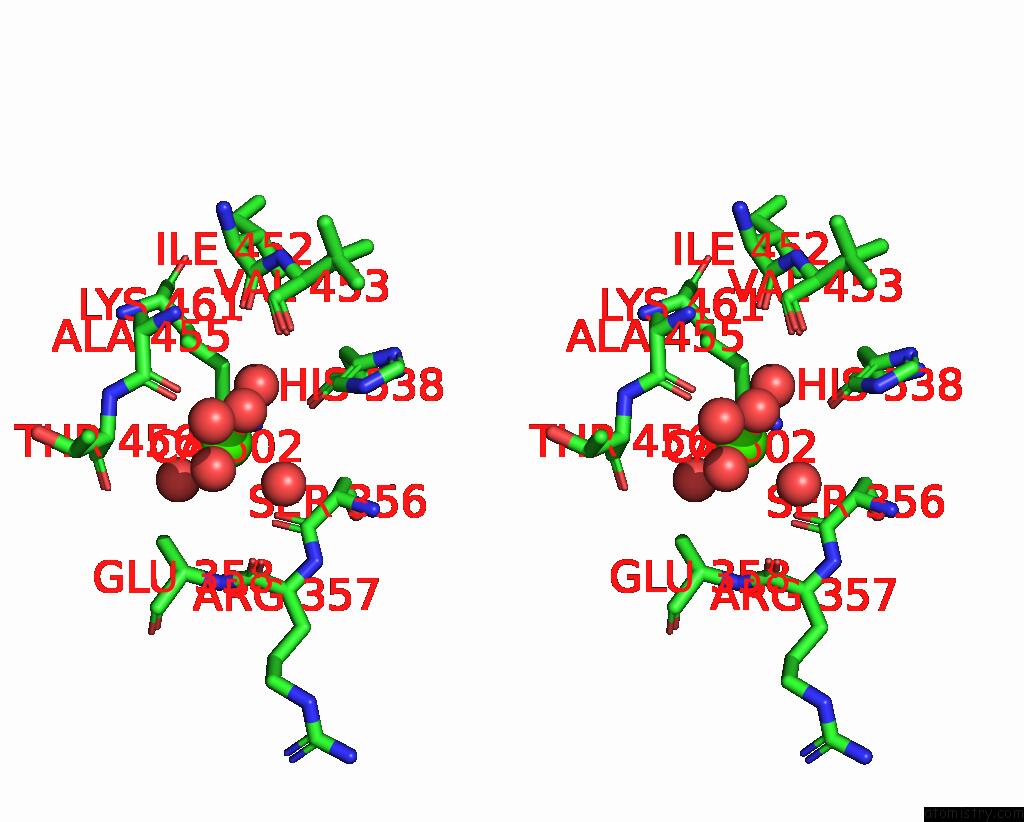
Stereo pair view
Mono view
Stereo pair view
A full contact list of Calcium with other atoms in the Ca binding
site number 2 of Human TRAP1 Nm in Complex with 42C within 5.0Å range:
|
Reference:
T.R.Stachowski,
S.Nithianantham,
M.Vanarotti,
K.Lopez,
M.Fischer.
Pan-HSP90 Ligand Binding Reveals Isoform-Specific Differences in Plasticity and Water Networks. Protein Sci. E4629 2023.
ISSN: ESSN 1469-896X
PubMed: 36938943
DOI: 10.1002/PRO.4629
Page generated: Thu Jul 10 01:44:35 2025
ISSN: ESSN 1469-896X
PubMed: 36938943
DOI: 10.1002/PRO.4629
Last articles
Fe in 2YXOFe in 2YRS
Fe in 2YXC
Fe in 2YNM
Fe in 2YVJ
Fe in 2YP1
Fe in 2YU2
Fe in 2YU1
Fe in 2YQB
Fe in 2YOO