Atomistry » Calcium » PDB 7ucy-7uwv » 7unp
Atomistry »
  Calcium »
    PDB 7ucy-7uwv »
      7unp »

Calcium in PDB 7unp: Crystal Structure of the Celr Catalytic Domain and CBM3C

Protein crystallography data

The structure of Crystal Structure of the Celr Catalytic Domain and CBM3C, PDB code: 7unp was solved by C.A.Bingman, N.Kuch, M.E.Kutsche, A.Parker, R.W.Smith, B.G.Fox, with X-Ray Crystallography technique. A brief refinement statistics is given in the table below:

Resolution Low / High (Å) 36.80 / 2.00
Space group P 1 21 1
Cell size a, b, c (Å), α, β, γ (°) 53.751, 91.076, 66.909, 90, 110.99, 90
R / Rfree (%) 16 / 20.7

Calcium Binding Sites:

The binding sites of Calcium atom in the Crystal Structure of the Celr Catalytic Domain and CBM3C (pdb code 7unp). This binding sites where shown within 5.0 Angstroms radius around Calcium atom.
In total 2 binding sites of Calcium where determined in the Crystal Structure of the Celr Catalytic Domain and CBM3C, PDB code: 7unp:
Jump to Calcium binding site number: 1; 2;

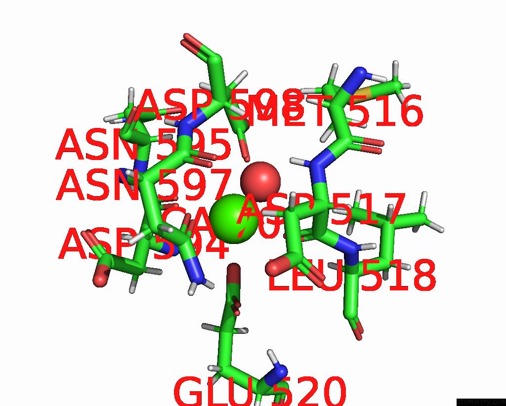
Calcium binding site 1 out of 2 in 7unp

Go back to Calcium Binding Sites List in 7unp
Calcium binding site 1 out of 2 in the Crystal Structure of the Celr Catalytic Domain and CBM3C


Mono view


Stereo pair view

A full contact list of Calcium with other atoms in the Ca binding site number 1 of Crystal Structure of the Celr Catalytic Domain and CBM3C within 5.0Å range:
probe atom residue distance (Å) B Occ
A:Ca701

b:22.4
occ:0.93
O A:ASP517 2.3 25.0 1.0
OE2 A:GLU520 2.3 26.7 1.0
OD1 A:ASN597 2.3 27.2 1.0
O A:ASP594 2.3 21.1 1.0
OD1 A:ASP598 2.4 18.3 1.0
O A:HOH808 2.5 21.9 1.0
CD A:GLU520 3.1 28.1 1.0
OE1 A:GLU520 3.3 28.6 1.0
HD21 A:ASN597 3.3 32.3 1.0
CG A:ASN597 3.3 30.5 1.0
CG A:ASP598 3.3 20.8 1.0
H A:ASP594 3.4 28.2 1.0
HB3 A:ASP594 3.4 30.9 1.0
C A:ASP517 3.4 20.4 1.0
C A:ASP594 3.4 20.0 1.0
H A:ASP517 3.5 23.5 1.0
HB3 A:ASP517 3.5 31.8 1.0
OD2 A:ASP598 3.6 17.3 1.0
ND2 A:ASN597 3.7 26.9 1.0
HA A:LEU518 4.0 28.3 1.0
HD23 A:LEU518 4.1 25.5 1.0
N A:ASP517 4.1 19.7 1.0
N A:ASP594 4.1 23.5 1.0
CA A:ASP594 4.1 24.2 1.0
CA A:ASP517 4.1 22.8 1.0
H A:ASN597 4.1 27.1 1.0
CB A:ASP594 4.2 25.8 1.0
HA A:ASN595 4.2 23.8 1.0
CB A:ASP517 4.3 26.5 1.0
HB3 A:MET516 4.4 27.9 1.0
N A:LEU518 4.5 20.5 1.0
N A:ASN595 4.5 17.9 1.0
HD22 A:ASN597 4.5 32.3 1.0
N A:ASP598 4.5 24.7 1.0
CG A:GLU520 4.6 30.4 1.0
C A:ASN597 4.6 25.8 1.0
CB A:ASP598 4.6 22.9 1.0
HA A:ASP598 4.6 26.0 1.0
CB A:ASN597 4.7 29.2 1.0
H A:ASP598 4.7 29.6 1.0
CA A:LEU518 4.7 23.5 1.0
CA A:ASN595 4.7 19.8 1.0
HG3 A:GLU520 4.7 36.5 1.0
N A:ASN597 4.8 22.6 1.0
CA A:ASP598 4.9 21.6 1.0
CG A:ASP594 4.9 27.3 1.0
HG2 A:GLU520 4.9 36.5 1.0
HB2 A:ASP594 4.9 30.9 1.0
O A:ASN597 4.9 23.5 1.0
CA A:ASN597 4.9 24.1 1.0
HB2 A:ASP598 5.0 27.4 1.0
H A:GLU520 5.0 34.6 1.0
C A:ASN595 5.0 25.0 1.0

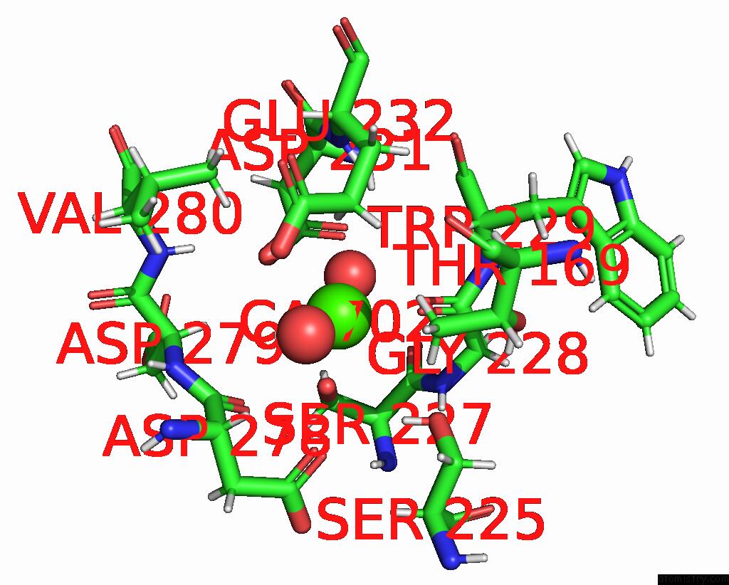
Calcium binding site 2 out of 2 in 7unp

Go back to Calcium Binding Sites List in 7unp
Calcium binding site 2 out of 2 in the Crystal Structure of the Celr Catalytic Domain and CBM3C


Mono view


Stereo pair view

A full contact list of Calcium with other atoms in the Ca binding site number 2 of Crystal Structure of the Celr Catalytic Domain and CBM3C within 5.0Å range:
probe atom residue distance (Å) B Occ
A:Ca702

b:33.2
occ:0.63
O A:GLY228 2.3 20.9 1.0
OG A:SER227 2.3 25.8 1.0
O A:HOH1021 2.3 29.7 1.0
OD1 A:ASP231 2.6 25.4 1.0
OD2 A:ASP231 2.8 20.3 1.0
CG A:ASP231 3.0 19.4 1.0
HG2 A:GLU232 3.2 20.6 1.0
C A:GLY228 3.4 23.8 1.0
CB A:SER227 3.5 23.4 1.0
HG A:SER225 3.6 35.2 1.0
HB2 A:SER227 3.6 28.1 1.0
O A:ASP278 3.6 18.7 1.0
N A:GLY228 3.8 25.0 1.0
C A:SER227 3.9 25.2 1.0
OD1 A:ASP278 3.9 30.4 1.0
HG23 A:THR169 3.9 21.1 1.0
HA A:ASP279 3.9 19.0 1.0
H A:GLY228 3.9 30.1 1.0
HG3 A:GLU232 4.0 20.6 1.0
CG A:GLU232 4.0 17.1 1.0
HA A:TRP229 4.1 26.4 1.0
C A:ASP278 4.1 18.5 1.0
OG A:SER225 4.1 29.4 1.0
H A:GLU232 4.2 19.8 1.0
O A:SER227 4.2 26.1 1.0
HA A:ASP278 4.2 23.2 1.0
HG21 A:THR169 4.2 21.1 1.0
CA A:GLY228 4.2 27.4 1.0
O A:HOH1093 4.2 32.1 1.0
HB3 A:SER227 4.2 28.1 1.0
CA A:SER227 4.3 25.8 1.0
HG22 A:VAL280 4.4 21.0 1.0
N A:TRP229 4.4 23.5 1.0
OE2 A:GLU232 4.4 25.8 1.0
H A:VAL280 4.4 15.9 1.0
H A:SER227 4.5 33.0 1.0
CG2 A:THR169 4.5 17.6 1.0
CB A:ASP231 4.5 21.2 1.0
H A:ASP231 4.6 19.4 1.0
N A:ASP279 4.6 16.4 1.0
CA A:TRP229 4.7 22.0 1.0
CA A:ASP279 4.7 15.8 1.0
CD A:GLU232 4.7 26.6 1.0
CA A:ASP278 4.7 19.3 1.0
N A:GLU232 4.8 16.4 1.0
HA3 A:GLY228 4.8 32.9 1.0
N A:SER227 4.8 27.5 1.0
HA2 A:GLY228 4.8 32.9 1.0
HB3 A:ASP231 4.9 25.4 1.0
CG A:ASP278 4.9 29.3 1.0
HG1 A:THR169 5.0 24.3 1.0
HB2 A:ASP231 5.0 25.4 1.0

Reference:

N.J.Kuch, M.E.Kutschke, A.Parker, C.A.Bingman, B.G.Fox. Contribution of Calcium Ligands in Substrate Binding and Product Release in the Acetovibrio Thermocellus Glycoside Hydrolase Family 9 Cellulase Celr. J.Biol.Chem. 04655 2023.
ISSN: ESSN 1083-351X
PubMed: 36990218
DOI: 10.1016/J.JBC.2023.104655
Page generated: Thu Jul 10 01:45:38 2025

Last articles

Fe in 2YXO
Fe in 2YRS
Fe in 2YXC
Fe in 2YNM
Fe in 2YVJ
Fe in 2YP1
Fe in 2YU2
Fe in 2YU1
Fe in 2YQB
Fe in 2YOO
© Copyright 2008-2020 by atomistry.com
Home   |    Site Map   |    Copyright   |    Contact us   |    Privacy