Calcium »
PDB 7vmk-7wa0 »
7w2v »
Calcium in PDB 7w2v: Crystal Structure of TXGH116 R786A Mutant From Thermoanaerobacterium Xylanolyticum with Glucose
Enzymatic activity of Crystal Structure of TXGH116 R786A Mutant From Thermoanaerobacterium Xylanolyticum with Glucose
All present enzymatic activity of Crystal Structure of TXGH116 R786A Mutant From Thermoanaerobacterium Xylanolyticum with Glucose:
3.2.1.45;
3.2.1.45;
Protein crystallography data
The structure of Crystal Structure of TXGH116 R786A Mutant From Thermoanaerobacterium Xylanolyticum with Glucose, PDB code: 7w2v
was solved by
M.Huang,
S.Pengthaisong,
R.Charoenwattanasatien,
J.Jitonnom,
J.R.Ketudatcairns,
with X-Ray Crystallography technique. A brief refinement statistics is given in the table below:
| Resolution Low / High (Å) | 30.00 / 1.80 |
| Space group | P 21 21 2 |
| Cell size a, b, c (Å), α, β, γ (°) | 177.444, 54.225, 83.112, 90, 90, 90 |
| R / Rfree (%) | 15.3 / 18.2 |
Calcium Binding Sites:
The binding sites of Calcium atom in the Crystal Structure of TXGH116 R786A Mutant From Thermoanaerobacterium Xylanolyticum with Glucose
(pdb code 7w2v). This binding sites where shown within
5.0 Angstroms radius around Calcium atom.
In total only one binding site of Calcium was determined in the Crystal Structure of TXGH116 R786A Mutant From Thermoanaerobacterium Xylanolyticum with Glucose, PDB code: 7w2v:
In total only one binding site of Calcium was determined in the Crystal Structure of TXGH116 R786A Mutant From Thermoanaerobacterium Xylanolyticum with Glucose, PDB code: 7w2v:
Calcium binding site 1 out of 1 in 7w2v
Go back to
Calcium binding site 1 out
of 1 in the Crystal Structure of TXGH116 R786A Mutant From Thermoanaerobacterium Xylanolyticum with Glucose
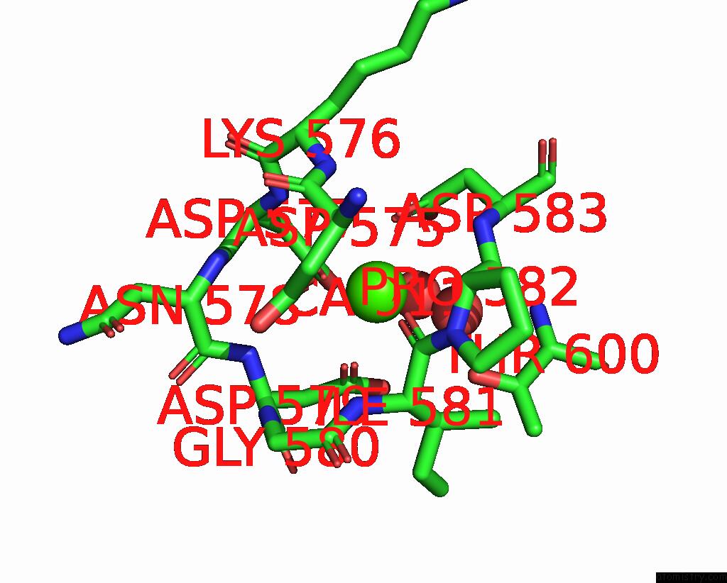
Mono view
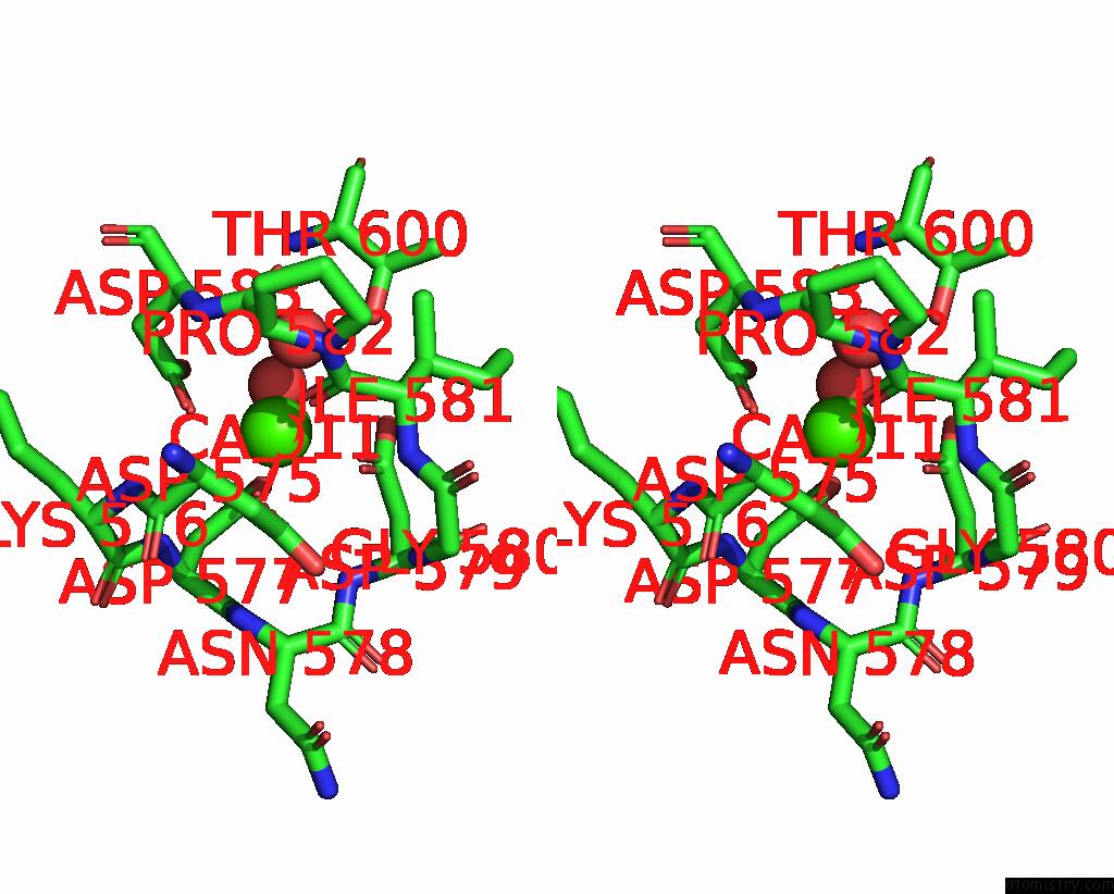
Stereo pair view
Mono view
Stereo pair view
A full contact list of Calcium with other atoms in the Ca binding
site number 1 of Crystal Structure of TXGH116 R786A Mutant From Thermoanaerobacterium Xylanolyticum with Glucose within 5.0Å range:
|
Reference:
M.Huang,
S.Pengthaisong,
R.Charoenwattanasatien,
N.Thinkumrob,
J.Jitonnom,
J.R.Ketudat Cairns.
Systematic Functional and Computational Analysis of Glucose-Binding Residues in Glycoside Hydrolase Family GH116. Catalysts V. 12 2022.
ISSN: ESSN 2073-4344
DOI: 10.3390/CATAL12030343
Page generated: Thu Jul 10 02:09:55 2025
ISSN: ESSN 2073-4344
DOI: 10.3390/CATAL12030343
Last articles
Fe in 2YXOFe in 2YRS
Fe in 2YXC
Fe in 2YNM
Fe in 2YVJ
Fe in 2YP1
Fe in 2YU2
Fe in 2YU1
Fe in 2YQB
Fe in 2YOO