Calcium »
PDB 7vmk-7wa0 »
7w51 »
Calcium in PDB 7w51: Crystal Structure of Fragmin Domain-1 in Complex with Actin (Adp-Form)
Protein crystallography data
The structure of Crystal Structure of Fragmin Domain-1 in Complex with Actin (Adp-Form), PDB code: 7w51
was solved by
S.Takeda,
with X-Ray Crystallography technique. A brief refinement statistics is given in the table below:
| Resolution Low / High (Å) | 30.31 / 1.20 |
| Space group | P 21 21 21 |
| Cell size a, b, c (Å), α, β, γ (°) | 57.22, 91.141, 115.24, 90, 90, 90 |
| R / Rfree (%) | 15.1 / 17.6 |
Other elements in 7w51:
The structure of Crystal Structure of Fragmin Domain-1 in Complex with Actin (Adp-Form) also contains other interesting chemical elements:
| Magnesium | (Mg) | 1 atom |
Calcium Binding Sites:
The binding sites of Calcium atom in the Crystal Structure of Fragmin Domain-1 in Complex with Actin (Adp-Form)
(pdb code 7w51). This binding sites where shown within
5.0 Angstroms radius around Calcium atom.
In total 2 binding sites of Calcium where determined in the Crystal Structure of Fragmin Domain-1 in Complex with Actin (Adp-Form), PDB code: 7w51:
Jump to Calcium binding site number: 1; 2;
In total 2 binding sites of Calcium where determined in the Crystal Structure of Fragmin Domain-1 in Complex with Actin (Adp-Form), PDB code: 7w51:
Jump to Calcium binding site number: 1; 2;
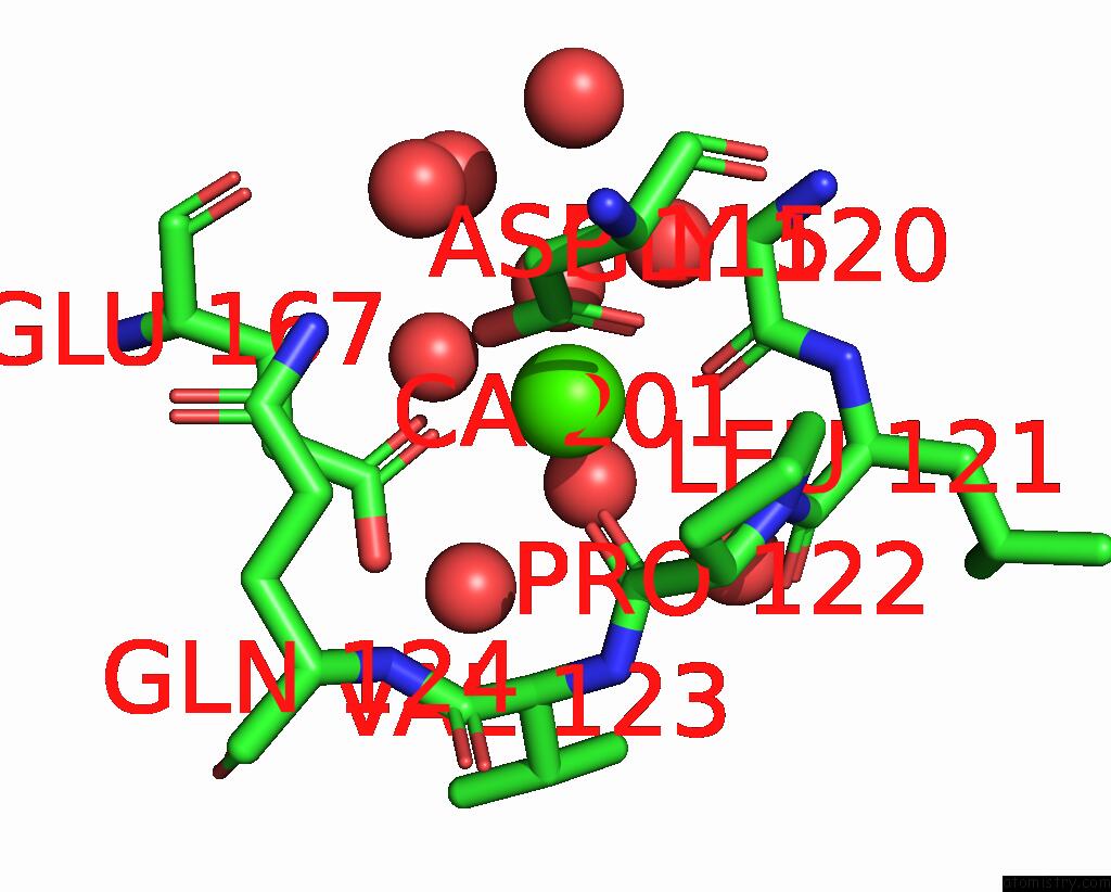
Calcium binding site 1 out of 2 in 7w51
Go back to
Calcium binding site 1 out
of 2 in the Crystal Structure of Fragmin Domain-1 in Complex with Actin (Adp-Form)
Mono view

Stereo pair view
Mono view
Stereo pair view
A full contact list of Calcium with other atoms in the Ca binding
site number 1 of Crystal Structure of Fragmin Domain-1 in Complex with Actin (Adp-Form) within 5.0Å range:
|
Calcium binding site 2 out of 2 in 7w51
Go back to
Calcium binding site 2 out
of 2 in the Crystal Structure of Fragmin Domain-1 in Complex with Actin (Adp-Form)

Mono view

Stereo pair view
Mono view
Stereo pair view
A full contact list of Calcium with other atoms in the Ca binding
site number 2 of Crystal Structure of Fragmin Domain-1 in Complex with Actin (Adp-Form) within 5.0Å range:
|
Reference:
Y.Kanematsu,
A.Narita,
T.Oda,
R.Koike,
M.Ota,
Y.Takano,
K.Moritsugu,
I.Fujiwara,
K.Tanaka,
H.Komatsu,
T.Nagae,
N.Watanabe,
M.Iwasa,
Y.Maeda,
S.Takeda.
Structures and Mechanisms of Actin Atp Hydrolysis. Proc.Natl.Acad.Sci.Usa V. 119 41119 2022.
ISSN: ESSN 1091-6490
PubMed: 36252034
DOI: 10.1073/PNAS.2122641119
Page generated: Thu Jul 10 02:11:30 2025
ISSN: ESSN 1091-6490
PubMed: 36252034
DOI: 10.1073/PNAS.2122641119
Last articles
F in 7QIRF in 7QI8
F in 7QC5
F in 7QG3
F in 7QHN
F in 7Q6P
F in 7QBZ
F in 7QC0
F in 7QAV
F in 7QA0