Calcium in PDB 7wb6: Crystal Structure of Bovine Pancreatic Trypsin in Complex with 4- Bromobenzamidine at Room Temperature
Enzymatic activity of Crystal Structure of Bovine Pancreatic Trypsin in Complex with 4- Bromobenzamidine at Room Temperature
All present enzymatic activity of Crystal Structure of Bovine Pancreatic Trypsin in Complex with 4- Bromobenzamidine at Room Temperature:
3.4.21.4;
3.4.21.4;
Protein crystallography data
The structure of Crystal Structure of Bovine Pancreatic Trypsin in Complex with 4- Bromobenzamidine at Room Temperature, PDB code: 7wb6
was solved by
N.Sakai,
H.Okumura,
M.Yamamoto,
T.Kumasaka,
with X-Ray Crystallography technique. A brief refinement statistics is given in the table below:
| Resolution Low / High (Å) | 44.07 / 1.48 |
| Space group | P 21 21 21 |
| Cell size a, b, c (Å), α, β, γ (°) | 54.57, 58.28, 67.35, 90, 90, 90 |
| R / Rfree (%) | 13.8 / 18.1 |
Other elements in 7wb6:
The structure of Crystal Structure of Bovine Pancreatic Trypsin in Complex with 4- Bromobenzamidine at Room Temperature also contains other interesting chemical elements:
| Bromine | (Br) | 1 atom |
Calcium Binding Sites:
The binding sites of Calcium atom in the Crystal Structure of Bovine Pancreatic Trypsin in Complex with 4- Bromobenzamidine at Room Temperature
(pdb code 7wb6). This binding sites where shown within
5.0 Angstroms radius around Calcium atom.
In total only one binding site of Calcium was determined in the Crystal Structure of Bovine Pancreatic Trypsin in Complex with 4- Bromobenzamidine at Room Temperature, PDB code: 7wb6:
In total only one binding site of Calcium was determined in the Crystal Structure of Bovine Pancreatic Trypsin in Complex with 4- Bromobenzamidine at Room Temperature, PDB code: 7wb6:
Calcium binding site 1 out of 1 in 7wb6
Go back to
Calcium binding site 1 out
of 1 in the Crystal Structure of Bovine Pancreatic Trypsin in Complex with 4- Bromobenzamidine at Room Temperature
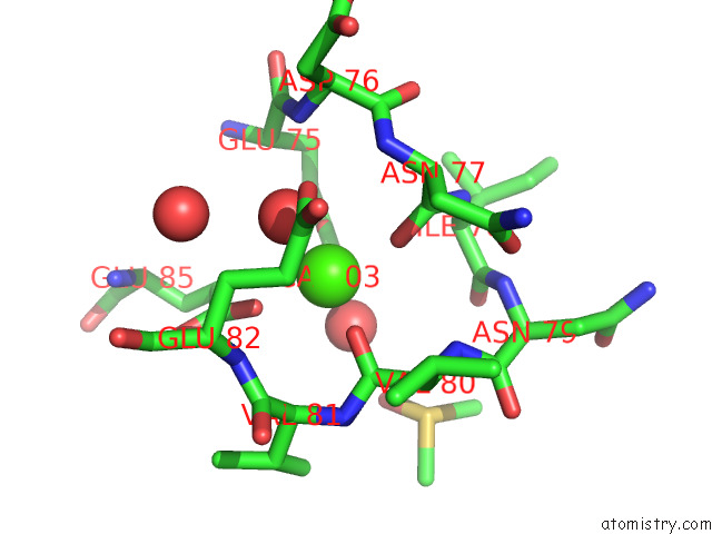
Mono view
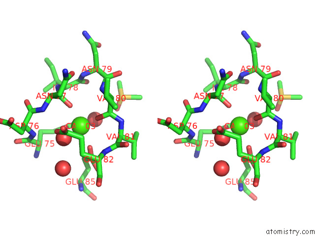
Stereo pair view
Mono view
Stereo pair view
A full contact list of Calcium with other atoms in the Ca binding
site number 1 of Crystal Structure of Bovine Pancreatic Trypsin in Complex with 4- Bromobenzamidine at Room Temperature within 5.0Å range:
|
Reference:
H.Okumura,
N.Sakai,
H.Murakami,
N.Mizuno,
Y.Nakamura,
G.Ueno,
T.Masunaga,
T.Kawamura,
S.Baba,
K.Hasegawa,
M.Yamamoto,
T.Kumasaka.
In Situ Crystal Data-Collection and Ligand-Screening System at Spring-8. Acta Crystallogr.,Sect.F V. 78 241 2022.
ISSN: ESSN 2053-230X
PubMed: 35647681
DOI: 10.1107/S2053230X22005283
Page generated: Fri Jul 19 05:39:48 2024
ISSN: ESSN 2053-230X
PubMed: 35647681
DOI: 10.1107/S2053230X22005283
Last articles
Zn in 9MJ5Zn in 9HNW
Zn in 9G0L
Zn in 9FNE
Zn in 9DZN
Zn in 9E0I
Zn in 9D32
Zn in 9DAK
Zn in 8ZXC
Zn in 8ZUF