Calcium »
PDB 7wa2-7wwo »
7wme »
Calcium in PDB 7wme: Crystal Structure of the Catalytic Domain of at-Higle
Protein crystallography data
The structure of Crystal Structure of the Catalytic Domain of at-Higle, PDB code: 7wme
was solved by
P.Verma,
P.Kumari,
S.Negi,
G.Yadav,
V.Gaur,
with X-Ray Crystallography technique. A brief refinement statistics is given in the table below:
| Resolution Low / High (Å) | 32.29 / 1.70 |
| Space group | P 21 21 21 |
| Cell size a, b, c (Å), α, β, γ (°) | 40.518, 53.452, 69.947, 90, 90, 90 |
| R / Rfree (%) | 16.4 / 19.4 |
Calcium Binding Sites:
The binding sites of Calcium atom in the Crystal Structure of the Catalytic Domain of at-Higle
(pdb code 7wme). This binding sites where shown within
5.0 Angstroms radius around Calcium atom.
In total only one binding site of Calcium was determined in the Crystal Structure of the Catalytic Domain of at-Higle, PDB code: 7wme:
In total only one binding site of Calcium was determined in the Crystal Structure of the Catalytic Domain of at-Higle, PDB code: 7wme:
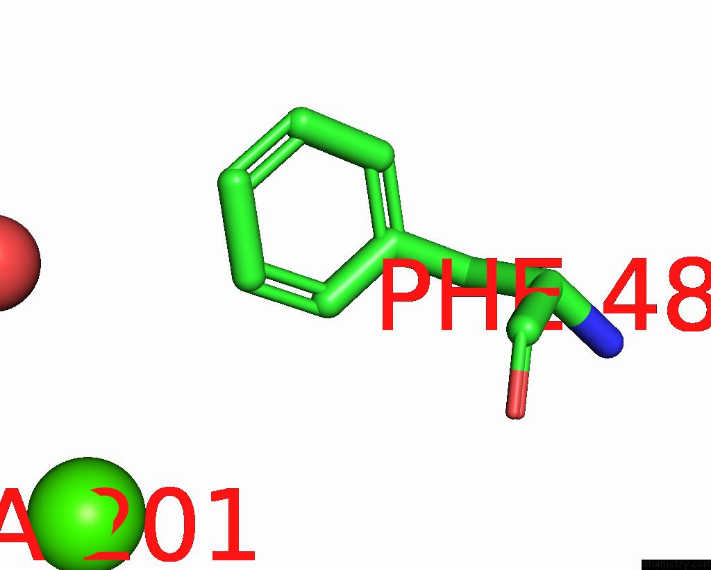
Calcium binding site 1 out of 1 in 7wme
Go back to
Calcium binding site 1 out
of 1 in the Crystal Structure of the Catalytic Domain of at-Higle
Mono view

Stereo pair view
Mono view
Stereo pair view
A full contact list of Calcium with other atoms in the Ca binding
site number 1 of Crystal Structure of the Catalytic Domain of at-Higle within 5.0Å range:
|
Reference:
P.Verma,
P.Kumari,
S.Negi,
G.Yadav,
V.Gaur.
Holliday Junction Resolution By at-Higle: An SLX1 Lineage Endonuclease From Arabidopsis Thaliana with A Novel in-Built Regulatory Mechanism. Nucleic Acids Res. V. 50 4630 2022.
ISSN: ESSN 1362-4962
PubMed: 35412622
DOI: 10.1093/NAR/GKAC239
Page generated: Thu Jul 10 02:17:18 2025
ISSN: ESSN 1362-4962
PubMed: 35412622
DOI: 10.1093/NAR/GKAC239
Last articles
Fe in 2YXOFe in 2YRS
Fe in 2YXC
Fe in 2YNM
Fe in 2YVJ
Fe in 2YP1
Fe in 2YU2
Fe in 2YU1
Fe in 2YQB
Fe in 2YOO