Calcium »
PDB 7wa2-7wwo »
7wmk »
Calcium in PDB 7wmk: Pqq-Dependent Alcohol Dehydrogenase Complexed with Pqq
Protein crystallography data
The structure of Pqq-Dependent Alcohol Dehydrogenase Complexed with Pqq, PDB code: 7wmk
was solved by
M.Chen,
H.Yang,
F.Lv,
with X-Ray Crystallography technique. A brief refinement statistics is given in the table below:
| Resolution Low / High (Å) | 44.15 / 1.47 |
| Space group | C 1 2 1 |
| Cell size a, b, c (Å), α, β, γ (°) | 109.973, 91.19, 57.332, 90, 103.78, 90 |
| R / Rfree (%) | 13.8 / 14 |
Other elements in 7wmk:
The structure of Pqq-Dependent Alcohol Dehydrogenase Complexed with Pqq also contains other interesting chemical elements:
| Sodium | (Na) | 1 atom |
| Chlorine | (Cl) | 2 atoms |
Calcium Binding Sites:
The binding sites of Calcium atom in the Pqq-Dependent Alcohol Dehydrogenase Complexed with Pqq
(pdb code 7wmk). This binding sites where shown within
5.0 Angstroms radius around Calcium atom.
In total 3 binding sites of Calcium where determined in the Pqq-Dependent Alcohol Dehydrogenase Complexed with Pqq, PDB code: 7wmk:
Jump to Calcium binding site number: 1; 2; 3;
In total 3 binding sites of Calcium where determined in the Pqq-Dependent Alcohol Dehydrogenase Complexed with Pqq, PDB code: 7wmk:
Jump to Calcium binding site number: 1; 2; 3;
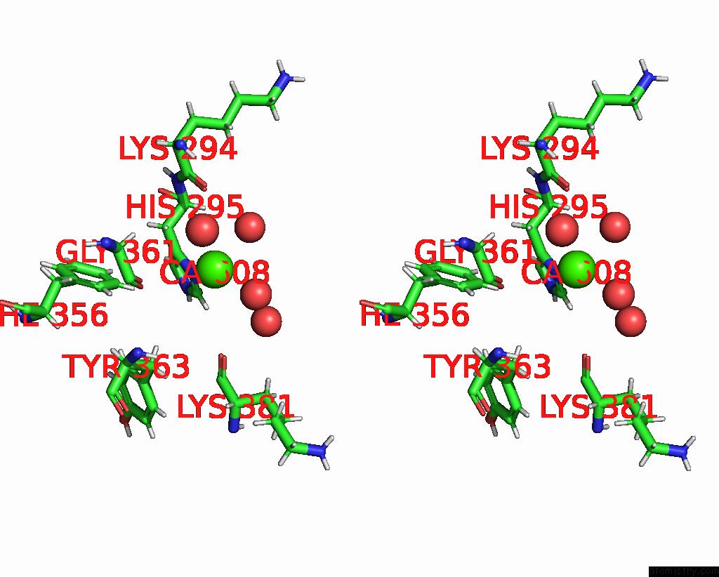
Calcium binding site 1 out of 3 in 7wmk
Go back to
Calcium binding site 1 out
of 3 in the Pqq-Dependent Alcohol Dehydrogenase Complexed with Pqq
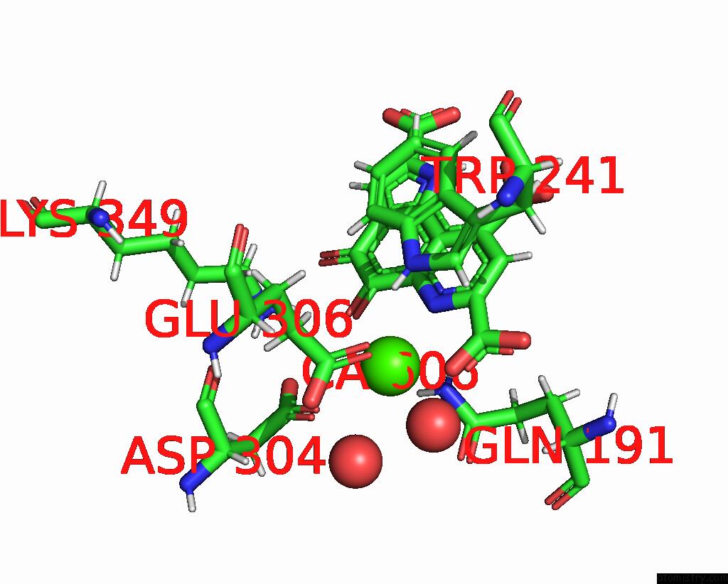
Mono view
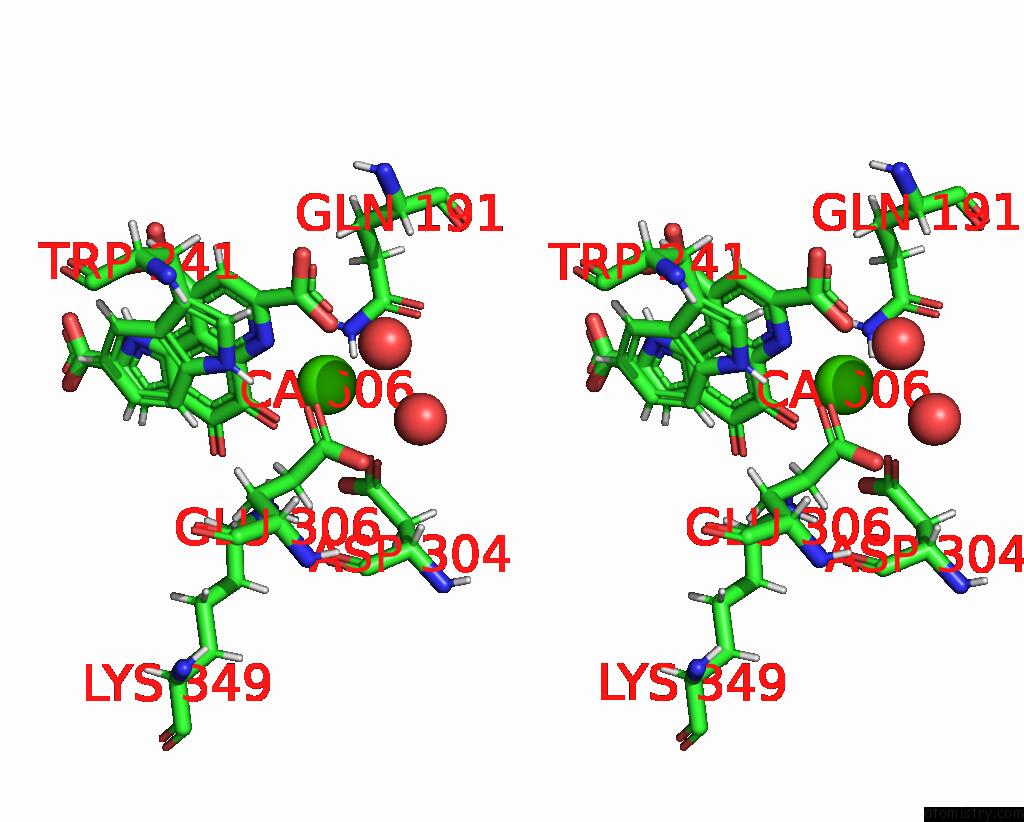
Stereo pair view
Mono view
Stereo pair view
A full contact list of Calcium with other atoms in the Ca binding
site number 1 of Pqq-Dependent Alcohol Dehydrogenase Complexed with Pqq within 5.0Å range:
|
Calcium binding site 2 out of 3 in 7wmk
Go back to
Calcium binding site 2 out
of 3 in the Pqq-Dependent Alcohol Dehydrogenase Complexed with Pqq
Mono view
Stereo pair view
Mono view
Stereo pair view
A full contact list of Calcium with other atoms in the Ca binding
site number 2 of Pqq-Dependent Alcohol Dehydrogenase Complexed with Pqq within 5.0Å range:
|
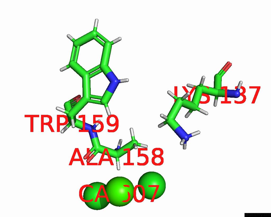
Calcium binding site 3 out of 3 in 7wmk
Go back to
Calcium binding site 3 out
of 3 in the Pqq-Dependent Alcohol Dehydrogenase Complexed with Pqq

Mono view
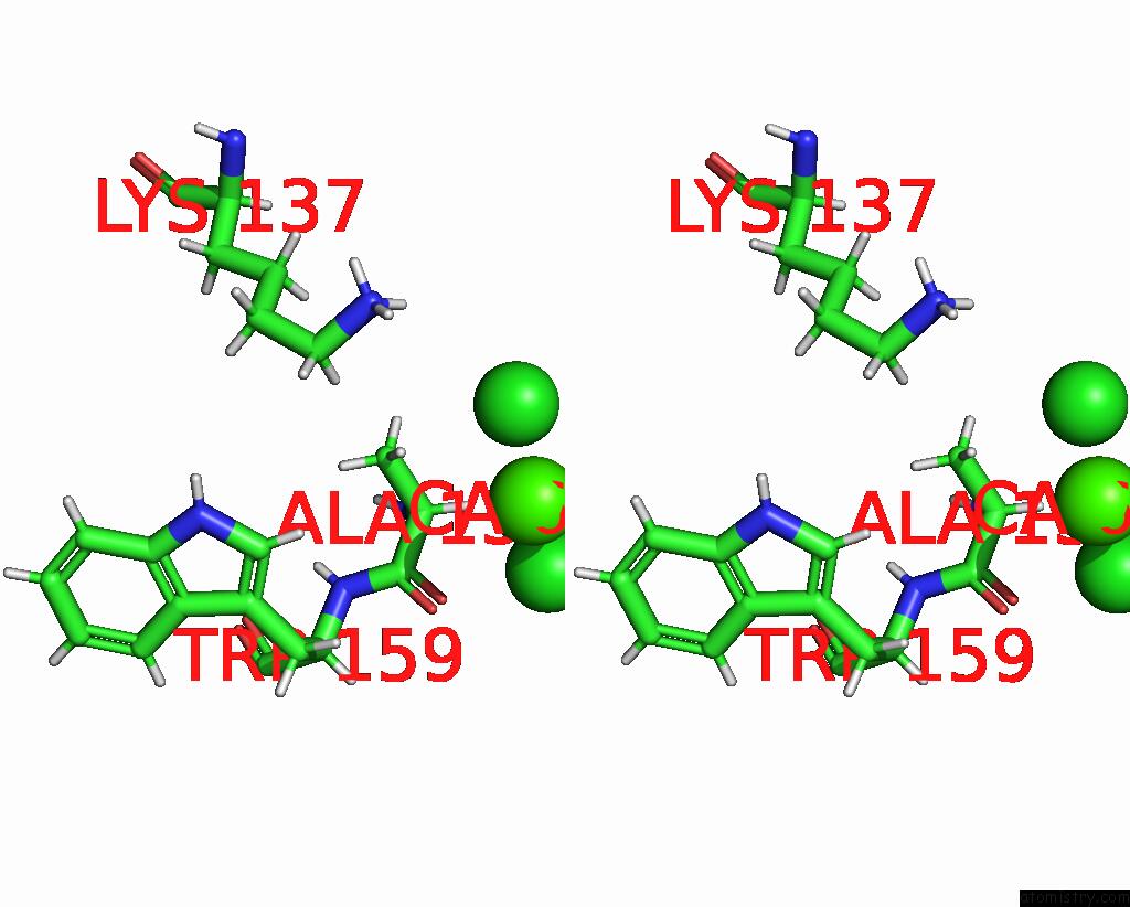
Stereo pair view
Mono view
Stereo pair view
A full contact list of Calcium with other atoms in the Ca binding
site number 3 of Pqq-Dependent Alcohol Dehydrogenase Complexed with Pqq within 5.0Å range:
|
Reference:
H.Yang,
R.Yan,
Y.Li,
Z.Lu,
X.Bie,
H.Zhao,
F.Lu,
M.Chen.
Structure-Function Analysis of A Quinone-Dependent Dehydrogenase Capable of Deoxynivalenol Detoxification. J.Agric.Food Chem. V. 70 6764 2022.
ISSN: ESSN 1520-5118
PubMed: 35613468
DOI: 10.1021/ACS.JAFC.2C01083
Page generated: Thu Jul 10 02:17:23 2025
ISSN: ESSN 1520-5118
PubMed: 35613468
DOI: 10.1021/ACS.JAFC.2C01083
Last articles
Fe in 2YXOFe in 2YRS
Fe in 2YXC
Fe in 2YNM
Fe in 2YVJ
Fe in 2YP1
Fe in 2YU2
Fe in 2YU1
Fe in 2YQB
Fe in 2YOO