Calcium »
PDB 7wxx-7xvv »
7x87 »
Calcium in PDB 7x87: The Complex Structure of Beta-1,2-Glucosyltransferase From Ignavibacterium Album with Sophotetraose Observed As Sophorose
Protein crystallography data
The structure of The Complex Structure of Beta-1,2-Glucosyltransferase From Ignavibacterium Album with Sophotetraose Observed As Sophorose, PDB code: 7x87
was solved by
K.Kobayashi,
H.Shimizu,
N.Tanaka,
K.Kuramochi,
H.Nakai,
M.Nakajima,
H.Taguchi,
with X-Ray Crystallography technique. A brief refinement statistics is given in the table below:
| Resolution Low / High (Å) | 47.91 / 1.79 |
| Space group | C 1 2 1 |
| Cell size a, b, c (Å), α, β, γ (°) | 165.135, 71.615, 130.308, 90, 104.89, 90 |
| R / Rfree (%) | 16.6 / 20.1 |
Calcium Binding Sites:
The binding sites of Calcium atom in the The Complex Structure of Beta-1,2-Glucosyltransferase From Ignavibacterium Album with Sophotetraose Observed As Sophorose
(pdb code 7x87). This binding sites where shown within
5.0 Angstroms radius around Calcium atom.
In total 4 binding sites of Calcium where determined in the The Complex Structure of Beta-1,2-Glucosyltransferase From Ignavibacterium Album with Sophotetraose Observed As Sophorose, PDB code: 7x87:
Jump to Calcium binding site number: 1; 2; 3; 4;
In total 4 binding sites of Calcium where determined in the The Complex Structure of Beta-1,2-Glucosyltransferase From Ignavibacterium Album with Sophotetraose Observed As Sophorose, PDB code: 7x87:
Jump to Calcium binding site number: 1; 2; 3; 4;
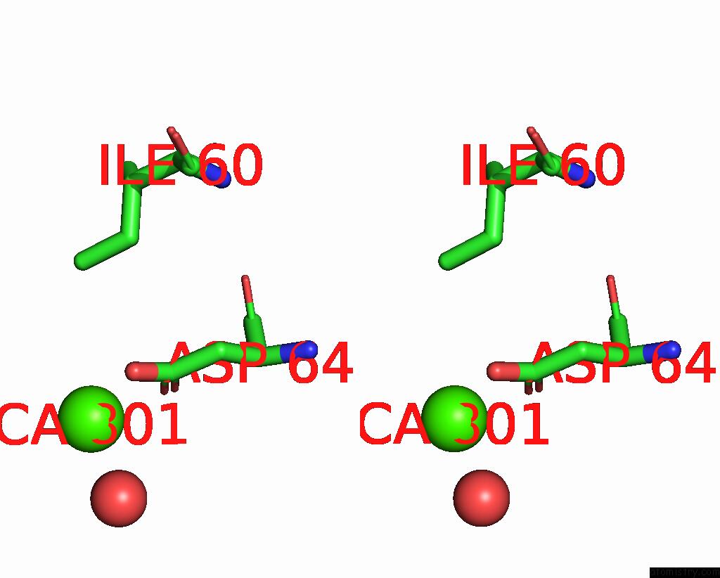
Calcium binding site 1 out of 4 in 7x87
Go back to
Calcium binding site 1 out
of 4 in the The Complex Structure of Beta-1,2-Glucosyltransferase From Ignavibacterium Album with Sophotetraose Observed As Sophorose

Mono view
Stereo pair view
Mono view
Stereo pair view
A full contact list of Calcium with other atoms in the Ca binding
site number 1 of The Complex Structure of Beta-1,2-Glucosyltransferase From Ignavibacterium Album with Sophotetraose Observed As Sophorose within 5.0Å range:
|
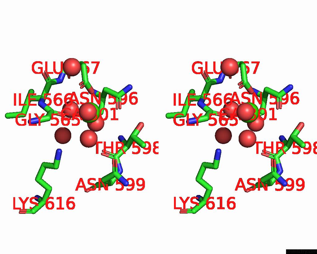
Calcium binding site 2 out of 4 in 7x87
Go back to
Calcium binding site 2 out
of 4 in the The Complex Structure of Beta-1,2-Glucosyltransferase From Ignavibacterium Album with Sophotetraose Observed As Sophorose

Mono view
Stereo pair view
Mono view
Stereo pair view
A full contact list of Calcium with other atoms in the Ca binding
site number 2 of The Complex Structure of Beta-1,2-Glucosyltransferase From Ignavibacterium Album with Sophotetraose Observed As Sophorose within 5.0Å range:
|
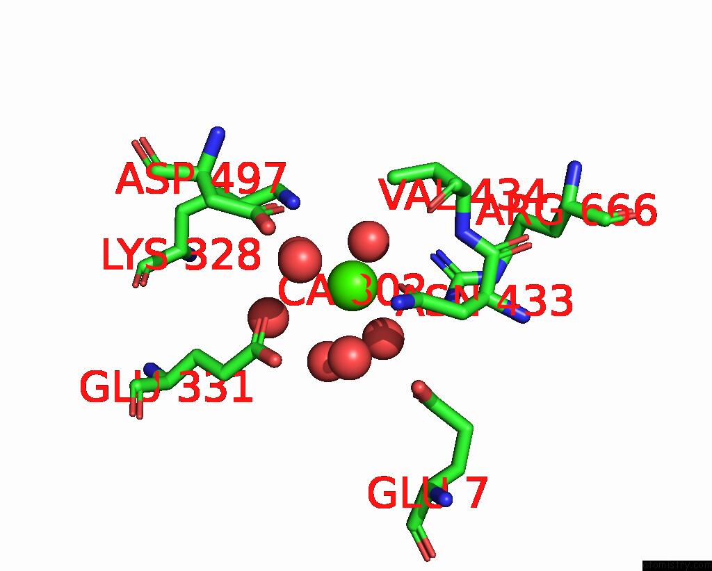
Calcium binding site 3 out of 4 in 7x87
Go back to
Calcium binding site 3 out
of 4 in the The Complex Structure of Beta-1,2-Glucosyltransferase From Ignavibacterium Album with Sophotetraose Observed As Sophorose
Mono view

Stereo pair view
Mono view
Stereo pair view
A full contact list of Calcium with other atoms in the Ca binding
site number 3 of The Complex Structure of Beta-1,2-Glucosyltransferase From Ignavibacterium Album with Sophotetraose Observed As Sophorose within 5.0Å range:
|
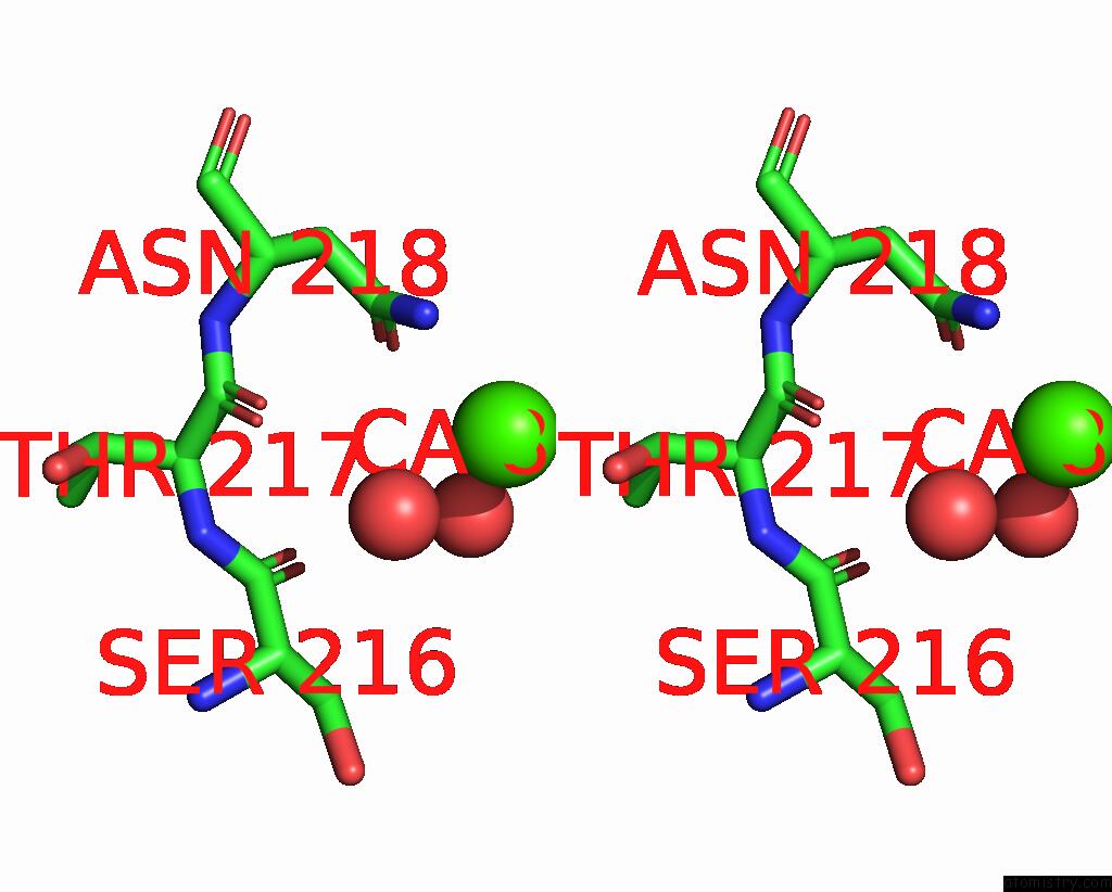
Calcium binding site 4 out of 4 in 7x87
Go back to
Calcium binding site 4 out
of 4 in the The Complex Structure of Beta-1,2-Glucosyltransferase From Ignavibacterium Album with Sophotetraose Observed As Sophorose

Mono view
Stereo pair view
Mono view
Stereo pair view
A full contact list of Calcium with other atoms in the Ca binding
site number 4 of The Complex Structure of Beta-1,2-Glucosyltransferase From Ignavibacterium Album with Sophotetraose Observed As Sophorose within 5.0Å range:
|
Reference:
K.Kobayashi,
H.Shimizu,
N.Tanaka,
K.Kuramochi,
H.Nakai,
M.Nakajima,
H.Taguchi.
Characterization and Structural Analyses of A Novel Glycosyltransferase Acting on the Beta-1,2-Glucosidic Linkages. J Biol Chem V. 298 01606 2022.
ISSN: ESSN 1083-351X
PubMed: 35065074
DOI: 10.1016/J.JBC.2022.101606
Page generated: Thu Jul 10 02:26:22 2025
ISSN: ESSN 1083-351X
PubMed: 35065074
DOI: 10.1016/J.JBC.2022.101606
Last articles
Fe in 2YXOFe in 2YRS
Fe in 2YXC
Fe in 2YNM
Fe in 2YVJ
Fe in 2YP1
Fe in 2YU2
Fe in 2YU1
Fe in 2YQB
Fe in 2YOO