Calcium »
PDB 7wxx-7xvv »
7xks »
Calcium in PDB 7xks: Crystal Structure of An Alkaline Pectate Lyase From Bacillus Clausii
Enzymatic activity of Crystal Structure of An Alkaline Pectate Lyase From Bacillus Clausii
All present enzymatic activity of Crystal Structure of An Alkaline Pectate Lyase From Bacillus Clausii:
4.2.2.2;
4.2.2.2;
Protein crystallography data
The structure of Crystal Structure of An Alkaline Pectate Lyase From Bacillus Clausii, PDB code: 7xks
was solved by
C.Zhou,
Y.Y.Zheng,
W.D.Liu,
Y.Ma,
with X-Ray Crystallography technique. A brief refinement statistics is given in the table below:
| Resolution Low / High (Å) | 27.27 / 1.78 |
| Space group | P 43 21 2 |
| Cell size a, b, c (Å), α, β, γ (°) | 109.085, 109.085, 44.977, 90, 90, 90 |
| R / Rfree (%) | 15.4 / 18.9 |
Calcium Binding Sites:
The binding sites of Calcium atom in the Crystal Structure of An Alkaline Pectate Lyase From Bacillus Clausii
(pdb code 7xks). This binding sites where shown within
5.0 Angstroms radius around Calcium atom.
In total only one binding site of Calcium was determined in the Crystal Structure of An Alkaline Pectate Lyase From Bacillus Clausii, PDB code: 7xks:
In total only one binding site of Calcium was determined in the Crystal Structure of An Alkaline Pectate Lyase From Bacillus Clausii, PDB code: 7xks:
Calcium binding site 1 out of 1 in 7xks
Go back to
Calcium binding site 1 out
of 1 in the Crystal Structure of An Alkaline Pectate Lyase From Bacillus Clausii
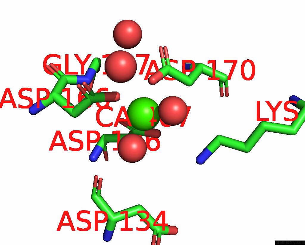
Mono view
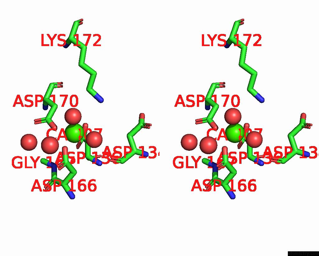
Stereo pair view
Mono view
Stereo pair view
A full contact list of Calcium with other atoms in the Ca binding
site number 1 of Crystal Structure of An Alkaline Pectate Lyase From Bacillus Clausii within 5.0Å range:
|
Reference:
C.Zhou,
Y.Cao,
Y.Xue,
W.Liu,
J.Ju,
Y.Ma.
Structure of An Alkaline Pectate Lyase and Rational Engineering with Improved Thermo-Alkaline Stability For Efficient Ramie Degumming. Int J Mol Sci V. 24 2022.
ISSN: ESSN 1422-0067
PubMed: 36613981
DOI: 10.3390/IJMS24010538
Page generated: Thu Jul 10 02:28:21 2025
ISSN: ESSN 1422-0067
PubMed: 36613981
DOI: 10.3390/IJMS24010538
Last articles
Fe in 2YXOFe in 2YRS
Fe in 2YXC
Fe in 2YNM
Fe in 2YVJ
Fe in 2YP1
Fe in 2YU2
Fe in 2YU1
Fe in 2YQB
Fe in 2YOO