Calcium »
PDB 7xvv-7yq7 »
7ygv »
Calcium in PDB 7ygv: Crystal Structure of the CA2+-Bound EFHD1/Swiprosin-2
Protein crystallography data
The structure of Crystal Structure of the CA2+-Bound EFHD1/Swiprosin-2, PDB code: 7ygv
was solved by
S.A.Mun,
J.Park,
J.Y.Kang,
T.Park,
M.Jin,
J.Ynag,
S.H.Eom,
with X-Ray Crystallography technique. A brief refinement statistics is given in the table below:
| Resolution Low / High (Å) | 31.72 / 2.80 |
| Space group | P 21 21 21 |
| Cell size a, b, c (Å), α, β, γ (°) | 44.251, 47.907, 63.434, 90, 90, 90 |
| R / Rfree (%) | 19.5 / 25.2 |
Other elements in 7ygv:
The structure of Crystal Structure of the CA2+-Bound EFHD1/Swiprosin-2 also contains other interesting chemical elements:
| Zinc | (Zn) | 1 atom |
Calcium Binding Sites:
The binding sites of Calcium atom in the Crystal Structure of the CA2+-Bound EFHD1/Swiprosin-2
(pdb code 7ygv). This binding sites where shown within
5.0 Angstroms radius around Calcium atom.
In total 2 binding sites of Calcium where determined in the Crystal Structure of the CA2+-Bound EFHD1/Swiprosin-2, PDB code: 7ygv:
Jump to Calcium binding site number: 1; 2;
In total 2 binding sites of Calcium where determined in the Crystal Structure of the CA2+-Bound EFHD1/Swiprosin-2, PDB code: 7ygv:
Jump to Calcium binding site number: 1; 2;
Calcium binding site 1 out of 2 in 7ygv
Go back to
Calcium binding site 1 out
of 2 in the Crystal Structure of the CA2+-Bound EFHD1/Swiprosin-2
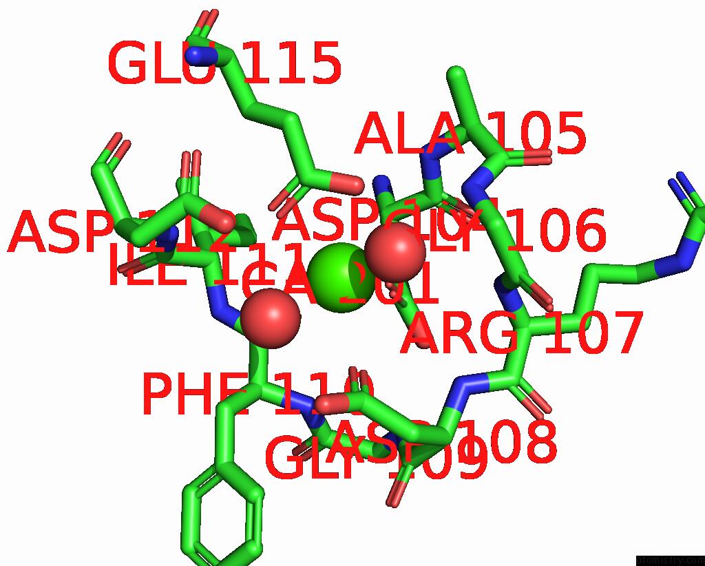
Mono view
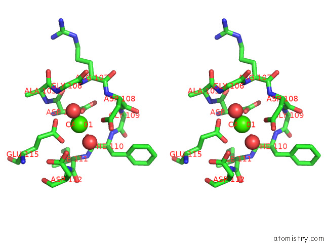
Stereo pair view
Mono view
Stereo pair view
A full contact list of Calcium with other atoms in the Ca binding
site number 1 of Crystal Structure of the CA2+-Bound EFHD1/Swiprosin-2 within 5.0Å range:
|
Calcium binding site 2 out of 2 in 7ygv
Go back to
Calcium binding site 2 out
of 2 in the Crystal Structure of the CA2+-Bound EFHD1/Swiprosin-2
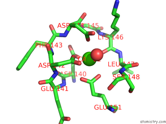
Mono view
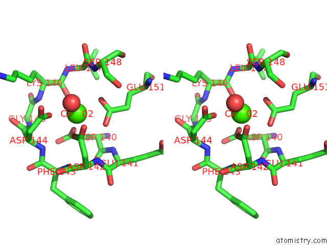
Stereo pair view
Mono view
Stereo pair view
A full contact list of Calcium with other atoms in the Ca binding
site number 2 of Crystal Structure of the CA2+-Bound EFHD1/Swiprosin-2 within 5.0Å range:
|
Reference:
S.A.Mun,
J.Park,
J.Y.Kang,
T.Park,
M.Jin,
J.Yang,
S.H.Eom.
Structural and Biochemical Insights Into Zn 2+ -Bound Ef-Hand Proteins, EFHD1 and EFHD2. Iucrj V. 10 233 2023.
ISSN: ESSN 2052-2525
PubMed: 36862489
DOI: 10.1107/S2052252523001501
Page generated: Fri Jul 19 06:13:54 2024
ISSN: ESSN 2052-2525
PubMed: 36862489
DOI: 10.1107/S2052252523001501
Last articles
Zn in 9JYWZn in 9IR4
Zn in 9IR3
Zn in 9GMX
Zn in 9GMW
Zn in 9JEJ
Zn in 9ERF
Zn in 9ERE
Zn in 9EGV
Zn in 9EGW