Calcium »
PDB 7xvv-7yq7 »
7ym0 »
Calcium in PDB 7ym0: Lysoplasmalogen-Specific Phospholipase D (Lypls-Pld) with CA2+
Protein crystallography data
The structure of Lysoplasmalogen-Specific Phospholipase D (Lypls-Pld) with CA2+, PDB code: 7ym0
was solved by
Y.Yasutake,
S.Sakasegawa,
D.Sugimori,
K.Murayama,
with X-Ray Crystallography technique. A brief refinement statistics is given in the table below:
| Resolution Low / High (Å) | 47.29 / 2.91 |
| Space group | P 43 21 2 |
| Cell size a, b, c (Å), α, β, γ (°) | 142.952, 142.952, 126.134, 90, 90, 90 |
| R / Rfree (%) | 17.7 / 21.5 |
Calcium Binding Sites:
The binding sites of Calcium atom in the Lysoplasmalogen-Specific Phospholipase D (Lypls-Pld) with CA2+
(pdb code 7ym0). This binding sites where shown within
5.0 Angstroms radius around Calcium atom.
In total 2 binding sites of Calcium where determined in the Lysoplasmalogen-Specific Phospholipase D (Lypls-Pld) with CA2+, PDB code: 7ym0:
Jump to Calcium binding site number: 1; 2;
In total 2 binding sites of Calcium where determined in the Lysoplasmalogen-Specific Phospholipase D (Lypls-Pld) with CA2+, PDB code: 7ym0:
Jump to Calcium binding site number: 1; 2;
Calcium binding site 1 out of 2 in 7ym0
Go back to
Calcium binding site 1 out
of 2 in the Lysoplasmalogen-Specific Phospholipase D (Lypls-Pld) with CA2+
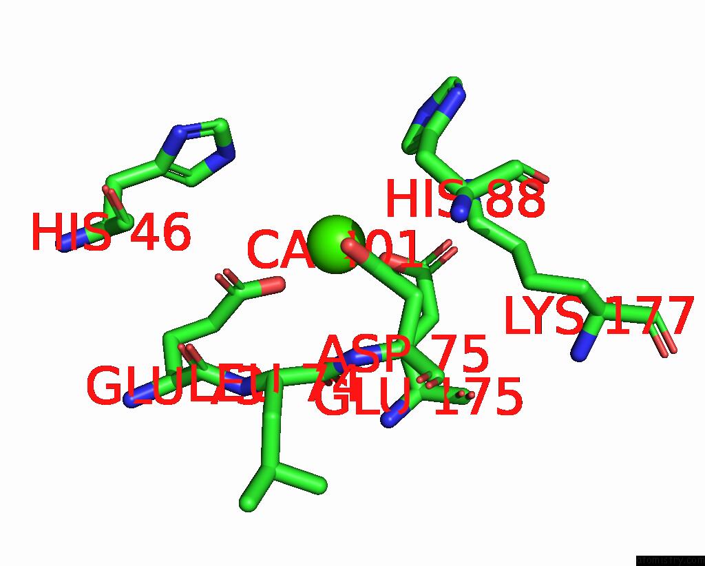
Mono view
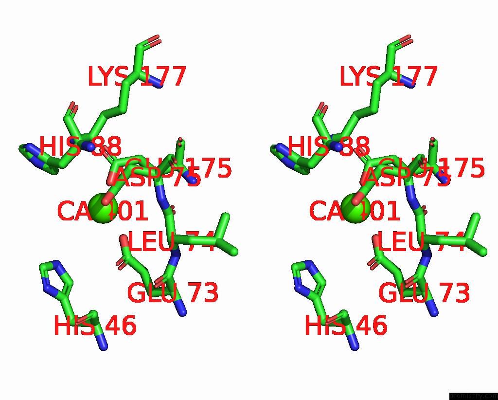
Stereo pair view
Mono view
Stereo pair view
A full contact list of Calcium with other atoms in the Ca binding
site number 1 of Lysoplasmalogen-Specific Phospholipase D (Lypls-Pld) with CA2+ within 5.0Å range:
|
Calcium binding site 2 out of 2 in 7ym0
Go back to
Calcium binding site 2 out
of 2 in the Lysoplasmalogen-Specific Phospholipase D (Lypls-Pld) with CA2+
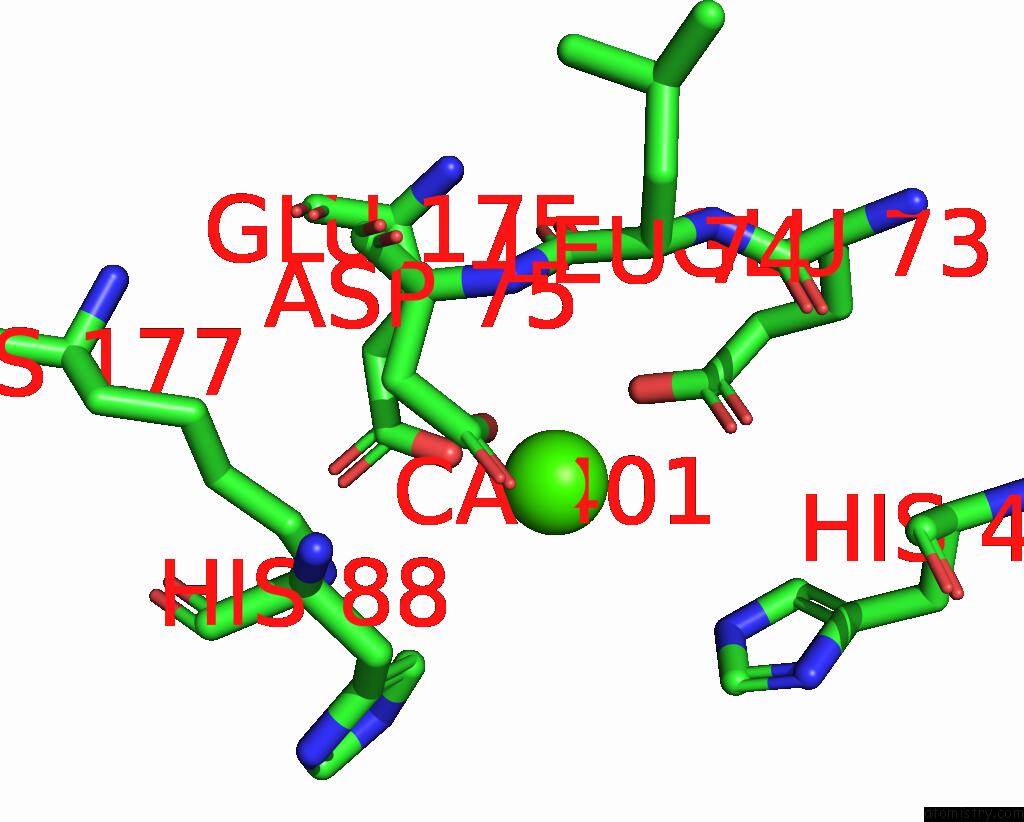
Mono view
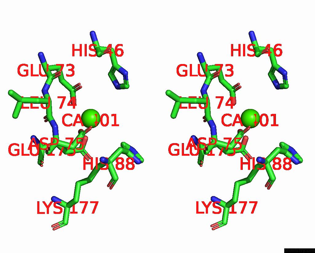
Stereo pair view
Mono view
Stereo pair view
A full contact list of Calcium with other atoms in the Ca binding
site number 2 of Lysoplasmalogen-Specific Phospholipase D (Lypls-Pld) with CA2+ within 5.0Å range:
|
Reference:
H.Hamana,
Y.Yasutake,
M.Kato-Murayama,
T.Hosaka,
M.Shirouzu,
S.I.Sakasegawa,
D.Sugimori,
K.Murayama.
Structural Basis For the Substrate Specificity Switching of Lysoplasmalogen-Specific Phospholipase D From Thermocrispum Sp. RD004668. Biosci.Biotechnol.Biochem. V. 87 74 2022.
ISSN: ISSN 0916-8451
PubMed: 36307380
DOI: 10.1093/BBB/ZBAC169
Page generated: Fri Jul 19 06:16:03 2024
ISSN: ISSN 0916-8451
PubMed: 36307380
DOI: 10.1093/BBB/ZBAC169
Last articles
Zn in 9JYWZn in 9IR4
Zn in 9IR3
Zn in 9GMX
Zn in 9GMW
Zn in 9JEJ
Zn in 9ERF
Zn in 9ERE
Zn in 9EGV
Zn in 9EGW