Calcium »
PDB 7ytq-7zci »
7z3j »
Calcium in PDB 7z3j: Structure of Crystallisable Rat Phospholipase C Gamma 1 in Complex with Inositol 1,4,5-Trisphosphate
Enzymatic activity of Structure of Crystallisable Rat Phospholipase C Gamma 1 in Complex with Inositol 1,4,5-Trisphosphate
All present enzymatic activity of Structure of Crystallisable Rat Phospholipase C Gamma 1 in Complex with Inositol 1,4,5-Trisphosphate:
3.1.4.11;
3.1.4.11;
Protein crystallography data
The structure of Structure of Crystallisable Rat Phospholipase C Gamma 1 in Complex with Inositol 1,4,5-Trisphosphate, PDB code: 7z3j
was solved by
N.Pinotsis,
T.D.Bunney,
M.Katan,
with X-Ray Crystallography technique. A brief refinement statistics is given in the table below:
| Resolution Low / High (Å) | 49.29 / 2.00 |
| Space group | P 21 21 21 |
| Cell size a, b, c (Å), α, β, γ (°) | 72.76, 82.44, 230.11, 90, 90, 90 |
| R / Rfree (%) | 22.4 / 25.6 |
Calcium Binding Sites:
The binding sites of Calcium atom in the Structure of Crystallisable Rat Phospholipase C Gamma 1 in Complex with Inositol 1,4,5-Trisphosphate
(pdb code 7z3j). This binding sites where shown within
5.0 Angstroms radius around Calcium atom.
In total 2 binding sites of Calcium where determined in the Structure of Crystallisable Rat Phospholipase C Gamma 1 in Complex with Inositol 1,4,5-Trisphosphate, PDB code: 7z3j:
Jump to Calcium binding site number: 1; 2;
In total 2 binding sites of Calcium where determined in the Structure of Crystallisable Rat Phospholipase C Gamma 1 in Complex with Inositol 1,4,5-Trisphosphate, PDB code: 7z3j:
Jump to Calcium binding site number: 1; 2;
Calcium binding site 1 out of 2 in 7z3j
Go back to
Calcium binding site 1 out
of 2 in the Structure of Crystallisable Rat Phospholipase C Gamma 1 in Complex with Inositol 1,4,5-Trisphosphate
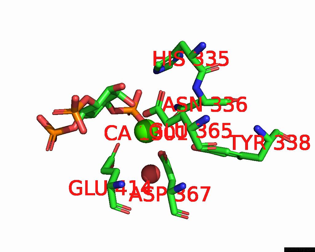
Mono view
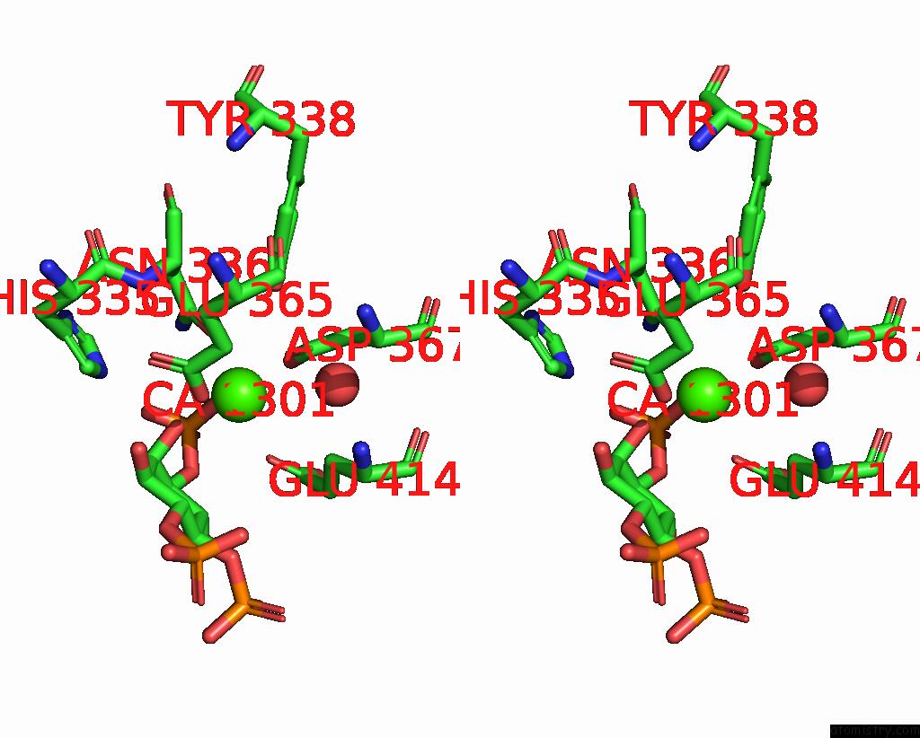
Stereo pair view
Mono view
Stereo pair view
A full contact list of Calcium with other atoms in the Ca binding
site number 1 of Structure of Crystallisable Rat Phospholipase C Gamma 1 in Complex with Inositol 1,4,5-Trisphosphate within 5.0Å range:
|
Calcium binding site 2 out of 2 in 7z3j
Go back to
Calcium binding site 2 out
of 2 in the Structure of Crystallisable Rat Phospholipase C Gamma 1 in Complex with Inositol 1,4,5-Trisphosphate
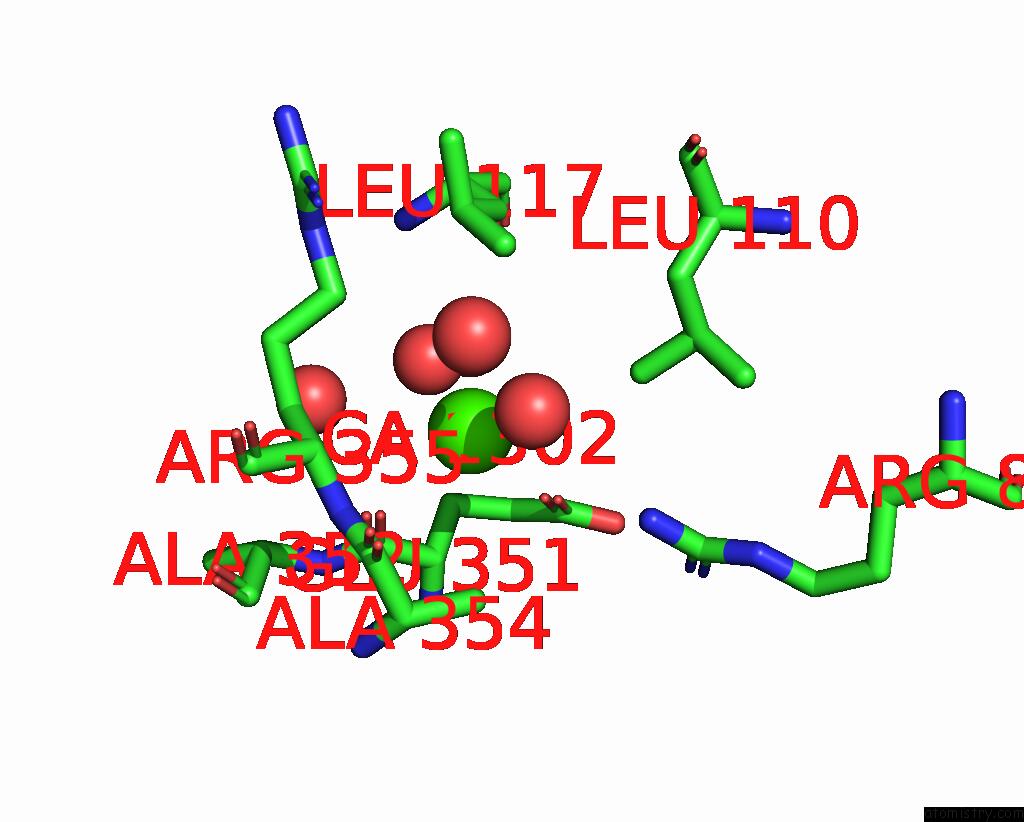
Mono view
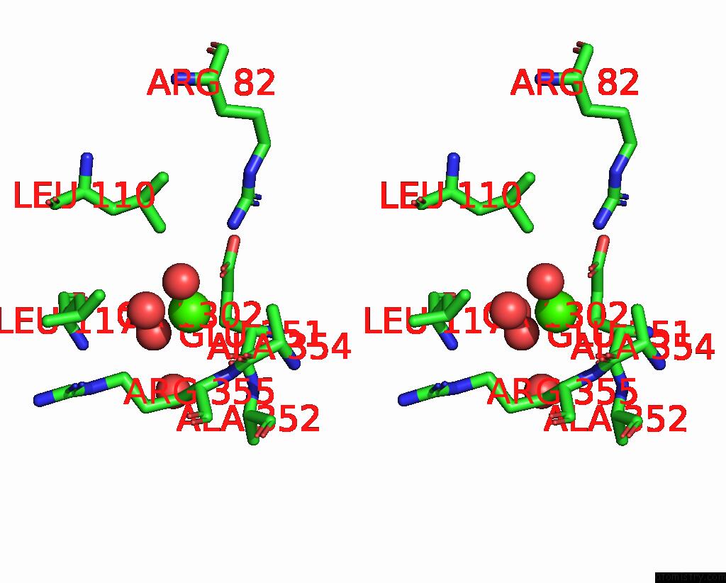
Stereo pair view
Mono view
Stereo pair view
A full contact list of Calcium with other atoms in the Ca binding
site number 2 of Structure of Crystallisable Rat Phospholipase C Gamma 1 in Complex with Inositol 1,4,5-Trisphosphate within 5.0Å range:
|
Reference:
K.I.P.Le Huray,
T.D.Bunney,
N.Pinotsis,
A.C.Kalli,
M.Katan.
Characterization of the Membrane Interactions of Phospholipase C Gamma Reveals Key Features of the Active Enzyme. Sci Adv V. 8 P9688 2022.
ISSN: ESSN 2375-2548
PubMed: 35749497
DOI: 10.1126/SCIADV.ABP9688
Page generated: Thu Jul 10 02:48:25 2025
ISSN: ESSN 2375-2548
PubMed: 35749497
DOI: 10.1126/SCIADV.ABP9688
Last articles
Fe in 2YXOFe in 2YRS
Fe in 2YXC
Fe in 2YNM
Fe in 2YVJ
Fe in 2YP1
Fe in 2YU2
Fe in 2YU1
Fe in 2YQB
Fe in 2YOO