Calcium »
PDB 8a2u-8awz »
8a5k »
Calcium in PDB 8a5k: Structural Analysis of 1-Deoxy-D-Xylulose 5-Phosphate Synthase From Pseudomonas Aeruginosa and Klebsiella Pneumoniae Reveals Conformational Changes Upon Cofactor Binding
Enzymatic activity of Structural Analysis of 1-Deoxy-D-Xylulose 5-Phosphate Synthase From Pseudomonas Aeruginosa and Klebsiella Pneumoniae Reveals Conformational Changes Upon Cofactor Binding
All present enzymatic activity of Structural Analysis of 1-Deoxy-D-Xylulose 5-Phosphate Synthase From Pseudomonas Aeruginosa and Klebsiella Pneumoniae Reveals Conformational Changes Upon Cofactor Binding:
2.2.1.7;
2.2.1.7;
Protein crystallography data
The structure of Structural Analysis of 1-Deoxy-D-Xylulose 5-Phosphate Synthase From Pseudomonas Aeruginosa and Klebsiella Pneumoniae Reveals Conformational Changes Upon Cofactor Binding, PDB code: 8a5k
was solved by
R.Hamid,
A.Hirsch,
with X-Ray Crystallography technique. A brief refinement statistics is given in the table below:
| Resolution Low / High (Å) | 46.92 / 2.30 |
| Space group | P 21 21 21 |
| Cell size a, b, c (Å), α, β, γ (°) | 116.819, 137.065, 231.445, 90, 90, 90 |
| R / Rfree (%) | 18.3 / 23.8 |
Other elements in 8a5k:
The structure of Structural Analysis of 1-Deoxy-D-Xylulose 5-Phosphate Synthase From Pseudomonas Aeruginosa and Klebsiella Pneumoniae Reveals Conformational Changes Upon Cofactor Binding also contains other interesting chemical elements:
| Magnesium | (Mg) | 6 atoms |
Calcium Binding Sites:
The binding sites of Calcium atom in the Structural Analysis of 1-Deoxy-D-Xylulose 5-Phosphate Synthase From Pseudomonas Aeruginosa and Klebsiella Pneumoniae Reveals Conformational Changes Upon Cofactor Binding
(pdb code 8a5k). This binding sites where shown within
5.0 Angstroms radius around Calcium atom.
In total 8 binding sites of Calcium where determined in the Structural Analysis of 1-Deoxy-D-Xylulose 5-Phosphate Synthase From Pseudomonas Aeruginosa and Klebsiella Pneumoniae Reveals Conformational Changes Upon Cofactor Binding, PDB code: 8a5k:
Jump to Calcium binding site number: 1; 2; 3; 4; 5; 6; 7; 8;
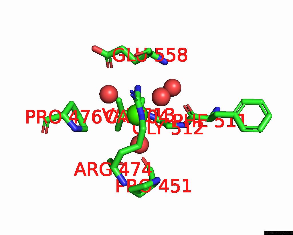
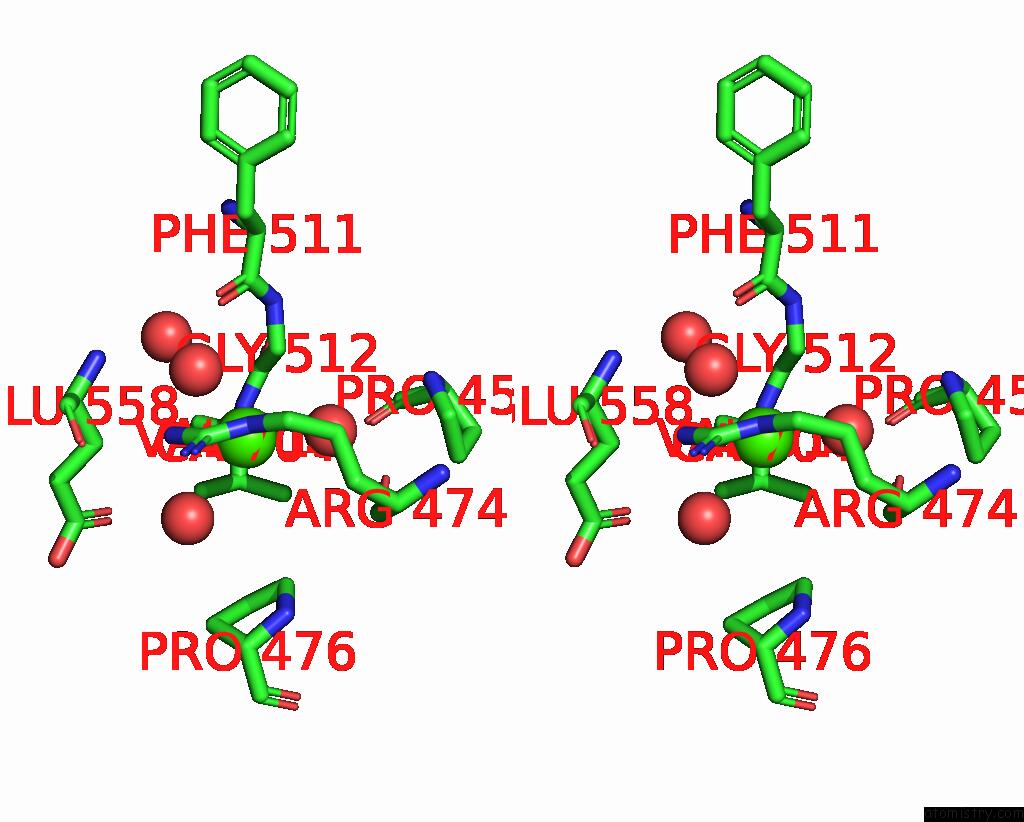
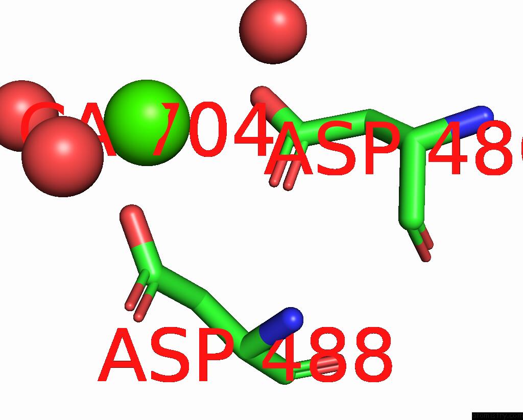
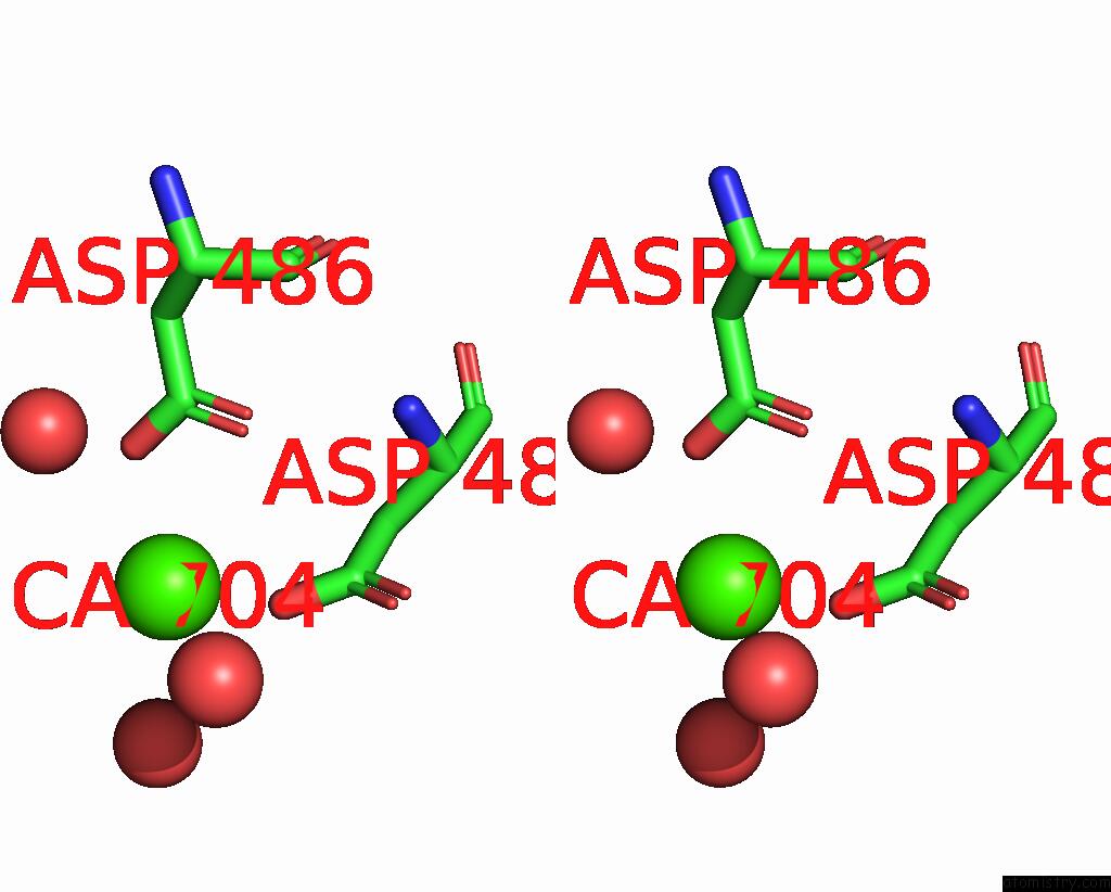
Calcium binding site 1 out of 8 in 8a5k
Go back to
Calcium binding site 1 out
of 8 in the Structural Analysis of 1-Deoxy-D-Xylulose 5-Phosphate Synthase From Pseudomonas Aeruginosa and Klebsiella Pneumoniae Reveals Conformational Changes Upon Cofactor Binding
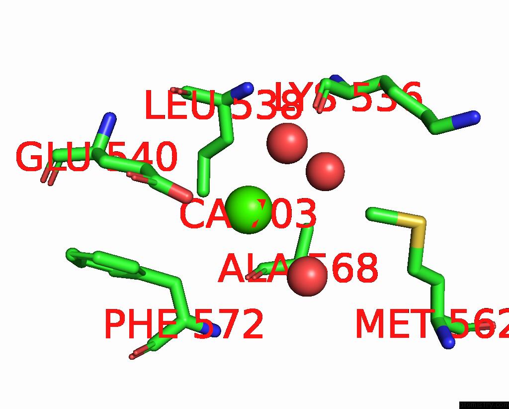
Mono view

Stereo pair view
Mono view
Stereo pair view
A full contact list of Calcium with other atoms in the Ca binding
site number 1 of Structural Analysis of 1-Deoxy-D-Xylulose 5-Phosphate Synthase From Pseudomonas Aeruginosa and Klebsiella Pneumoniae Reveals Conformational Changes Upon Cofactor Binding within 5.0Å range:
|
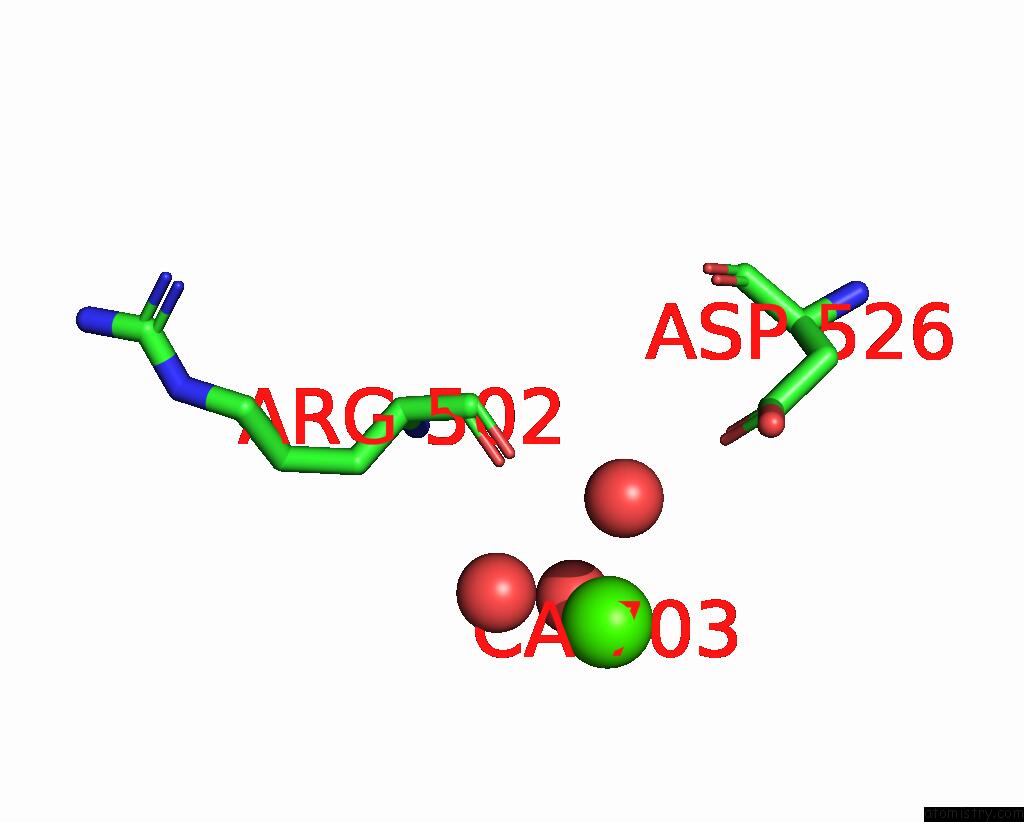
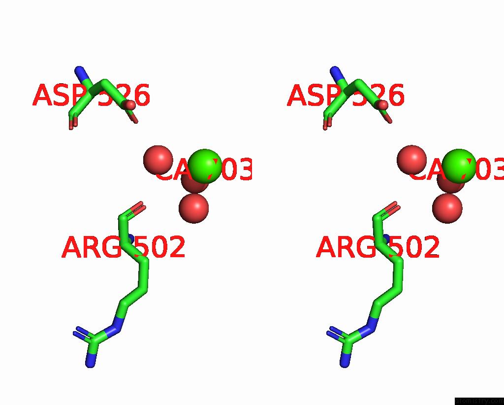
Calcium binding site 2 out of 8 in 8a5k
Go back to
Calcium binding site 2 out
of 8 in the Structural Analysis of 1-Deoxy-D-Xylulose 5-Phosphate Synthase From Pseudomonas Aeruginosa and Klebsiella Pneumoniae Reveals Conformational Changes Upon Cofactor Binding
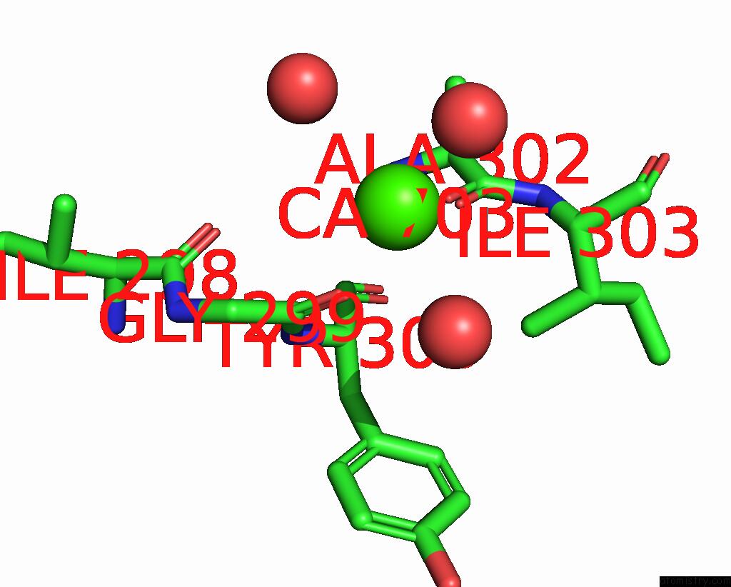
Mono view
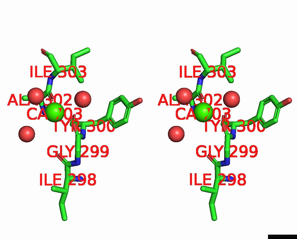
Stereo pair view
Mono view
Stereo pair view
A full contact list of Calcium with other atoms in the Ca binding
site number 2 of Structural Analysis of 1-Deoxy-D-Xylulose 5-Phosphate Synthase From Pseudomonas Aeruginosa and Klebsiella Pneumoniae Reveals Conformational Changes Upon Cofactor Binding within 5.0Å range:
|
Calcium binding site 3 out of 8 in 8a5k
Go back to
Calcium binding site 3 out
of 8 in the Structural Analysis of 1-Deoxy-D-Xylulose 5-Phosphate Synthase From Pseudomonas Aeruginosa and Klebsiella Pneumoniae Reveals Conformational Changes Upon Cofactor Binding
Mono view

Stereo pair view
Mono view
Stereo pair view
A full contact list of Calcium with other atoms in the Ca binding
site number 3 of Structural Analysis of 1-Deoxy-D-Xylulose 5-Phosphate Synthase From Pseudomonas Aeruginosa and Klebsiella Pneumoniae Reveals Conformational Changes Upon Cofactor Binding within 5.0Å range:
|
Calcium binding site 4 out of 8 in 8a5k
Go back to
Calcium binding site 4 out
of 8 in the Structural Analysis of 1-Deoxy-D-Xylulose 5-Phosphate Synthase From Pseudomonas Aeruginosa and Klebsiella Pneumoniae Reveals Conformational Changes Upon Cofactor Binding
Mono view
Stereo pair view
Mono view
Stereo pair view
A full contact list of Calcium with other atoms in the Ca binding
site number 4 of Structural Analysis of 1-Deoxy-D-Xylulose 5-Phosphate Synthase From Pseudomonas Aeruginosa and Klebsiella Pneumoniae Reveals Conformational Changes Upon Cofactor Binding within 5.0Å range:
|
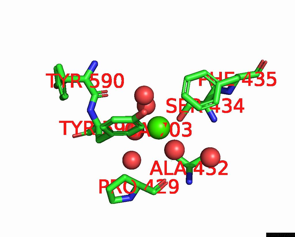
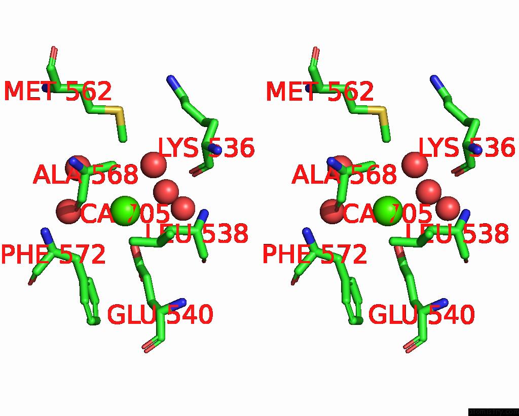
Calcium binding site 5 out of 8 in 8a5k
Go back to
Calcium binding site 5 out
of 8 in the Structural Analysis of 1-Deoxy-D-Xylulose 5-Phosphate Synthase From Pseudomonas Aeruginosa and Klebsiella Pneumoniae Reveals Conformational Changes Upon Cofactor Binding
Mono view
Stereo pair view
Mono view
Stereo pair view
A full contact list of Calcium with other atoms in the Ca binding
site number 5 of Structural Analysis of 1-Deoxy-D-Xylulose 5-Phosphate Synthase From Pseudomonas Aeruginosa and Klebsiella Pneumoniae Reveals Conformational Changes Upon Cofactor Binding within 5.0Å range:
|
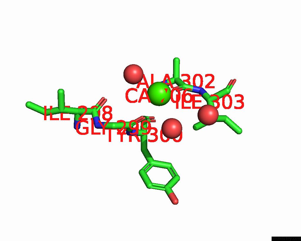
Calcium binding site 6 out of 8 in 8a5k
Go back to
Calcium binding site 6 out
of 8 in the Structural Analysis of 1-Deoxy-D-Xylulose 5-Phosphate Synthase From Pseudomonas Aeruginosa and Klebsiella Pneumoniae Reveals Conformational Changes Upon Cofactor Binding
Mono view
Stereo pair view
Mono view
Stereo pair view
A full contact list of Calcium with other atoms in the Ca binding
site number 6 of Structural Analysis of 1-Deoxy-D-Xylulose 5-Phosphate Synthase From Pseudomonas Aeruginosa and Klebsiella Pneumoniae Reveals Conformational Changes Upon Cofactor Binding within 5.0Å range:
|
Calcium binding site 7 out of 8 in 8a5k
Go back to
Calcium binding site 7 out
of 8 in the Structural Analysis of 1-Deoxy-D-Xylulose 5-Phosphate Synthase From Pseudomonas Aeruginosa and Klebsiella Pneumoniae Reveals Conformational Changes Upon Cofactor Binding

Mono view
Stereo pair view
Mono view
Stereo pair view
A full contact list of Calcium with other atoms in the Ca binding
site number 7 of Structural Analysis of 1-Deoxy-D-Xylulose 5-Phosphate Synthase From Pseudomonas Aeruginosa and Klebsiella Pneumoniae Reveals Conformational Changes Upon Cofactor Binding within 5.0Å range:
|
Calcium binding site 8 out of 8 in 8a5k
Go back to
Calcium binding site 8 out
of 8 in the Structural Analysis of 1-Deoxy-D-Xylulose 5-Phosphate Synthase From Pseudomonas Aeruginosa and Klebsiella Pneumoniae Reveals Conformational Changes Upon Cofactor Binding
Mono view

Stereo pair view
Mono view
Stereo pair view
A full contact list of Calcium with other atoms in the Ca binding
site number 8 of Structural Analysis of 1-Deoxy-D-Xylulose 5-Phosphate Synthase From Pseudomonas Aeruginosa and Klebsiella Pneumoniae Reveals Conformational Changes Upon Cofactor Binding within 5.0Å range:
|
Reference:
R.Hamid,
S.Adam,
A.Lacour,
L.Monjas,
A.Hirsch.
Structural Analysis of 1-Deoxy-D-Xylulose 5-Phosphate Synthase From Pseudomonas Aeruginosa and Klebsiella Pneumoniae Reveals Conformational Changes Upon Cofactor Binding To Be Published.
Page generated: Thu Jul 10 03:04:12 2025
Last articles
Cl in 5G3TCl in 5G54
Cl in 5G4A
Cl in 5G4Q
Cl in 5G47
Cl in 5G42
Cl in 5G3S
Cl in 5G2P
Cl in 5G2T
Cl in 5G36