Calcium »
PDB 8a2u-8awz »
8a8d »
Calcium in PDB 8a8d: Atp Sulfurylase From Methanothermococcus Thermolithotrophicus - Monoclinic Form
Enzymatic activity of Atp Sulfurylase From Methanothermococcus Thermolithotrophicus - Monoclinic Form
All present enzymatic activity of Atp Sulfurylase From Methanothermococcus Thermolithotrophicus - Monoclinic Form:
2.7.7.4;
2.7.7.4;
Protein crystallography data
The structure of Atp Sulfurylase From Methanothermococcus Thermolithotrophicus - Monoclinic Form, PDB code: 8a8d
was solved by
M.Jespersen,
T.Wagner,
with X-Ray Crystallography technique. A brief refinement statistics is given in the table below:
| Resolution Low / High (Å) | 52.28 / 2.10 |
| Space group | C 1 2 1 |
| Cell size a, b, c (Å), α, β, γ (°) | 185.282, 54.525, 85.43, 90, 95.91, 90 |
| R / Rfree (%) | 19.9 / 23.9 |
Other elements in 8a8d:
The structure of Atp Sulfurylase From Methanothermococcus Thermolithotrophicus - Monoclinic Form also contains other interesting chemical elements:
| Zinc | (Zn) | 2 atoms |
Calcium Binding Sites:
The binding sites of Calcium atom in the Atp Sulfurylase From Methanothermococcus Thermolithotrophicus - Monoclinic Form
(pdb code 8a8d). This binding sites where shown within
5.0 Angstroms radius around Calcium atom.
In total 3 binding sites of Calcium where determined in the Atp Sulfurylase From Methanothermococcus Thermolithotrophicus - Monoclinic Form, PDB code: 8a8d:
Jump to Calcium binding site number: 1; 2; 3;
In total 3 binding sites of Calcium where determined in the Atp Sulfurylase From Methanothermococcus Thermolithotrophicus - Monoclinic Form, PDB code: 8a8d:
Jump to Calcium binding site number: 1; 2; 3;
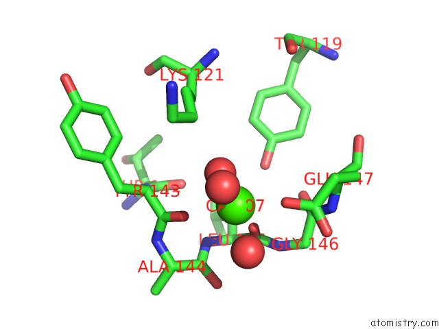
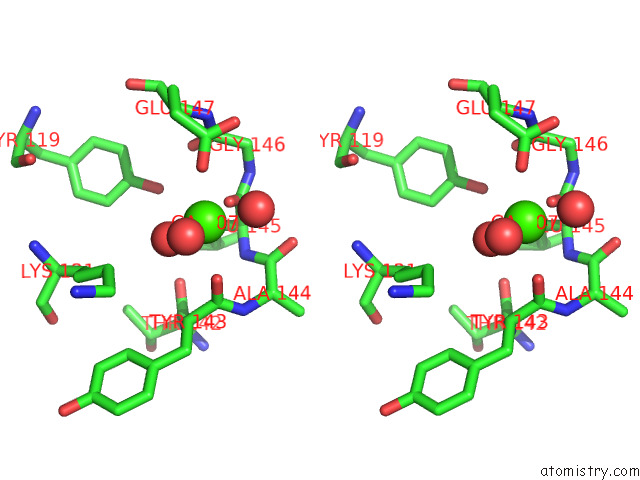
Calcium binding site 1 out of 3 in 8a8d
Go back to
Calcium binding site 1 out
of 3 in the Atp Sulfurylase From Methanothermococcus Thermolithotrophicus - Monoclinic Form
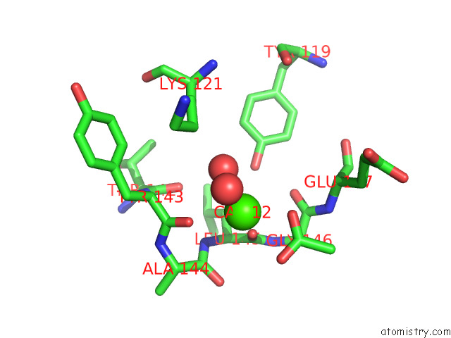
Mono view
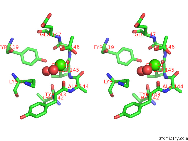
Stereo pair view
Mono view
Stereo pair view
A full contact list of Calcium with other atoms in the Ca binding
site number 1 of Atp Sulfurylase From Methanothermococcus Thermolithotrophicus - Monoclinic Form within 5.0Å range:
|
Calcium binding site 2 out of 3 in 8a8d
Go back to
Calcium binding site 2 out
of 3 in the Atp Sulfurylase From Methanothermococcus Thermolithotrophicus - Monoclinic Form
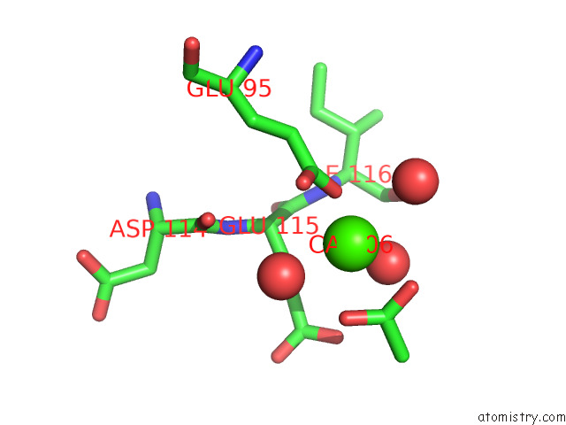
Mono view
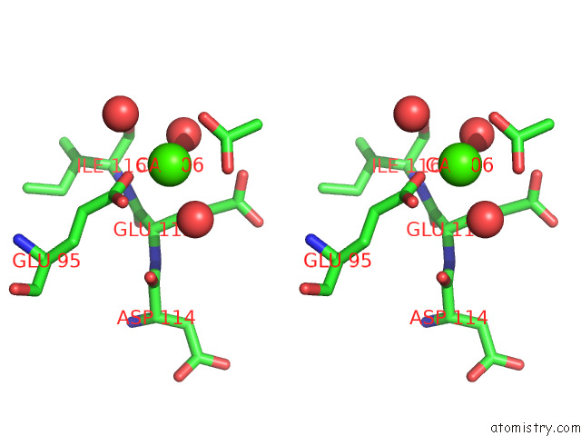
Stereo pair view
Mono view
Stereo pair view
A full contact list of Calcium with other atoms in the Ca binding
site number 2 of Atp Sulfurylase From Methanothermococcus Thermolithotrophicus - Monoclinic Form within 5.0Å range:
|
Calcium binding site 3 out of 3 in 8a8d
Go back to
Calcium binding site 3 out
of 3 in the Atp Sulfurylase From Methanothermococcus Thermolithotrophicus - Monoclinic Form
Mono view
Stereo pair view
Mono view
Stereo pair view
A full contact list of Calcium with other atoms in the Ca binding
site number 3 of Atp Sulfurylase From Methanothermococcus Thermolithotrophicus - Monoclinic Form within 5.0Å range:
|
Reference:
M.Jespersen,
T.Wagner.
How A Methanogen Assimilates Sulfate: Structural and Functional Elucidation of the Complete Sulfate-Reduction Pathway. To Be Published.
Page generated: Thu Jul 10 03:05:44 2025
Last articles
F in 4D6TF in 4D5H
F in 4D42
F in 4D43
F in 4D4V
F in 4D3B
F in 4D3A
F in 4D38
F in 4D37
F in 4D33