Calcium »
PDB 8ax0-8bad »
8b0v »
Calcium in PDB 8b0v: Crystal Structure of C-Terminal Domain of Pseudomonas Aeruginosa Lexa G91D Mutant
Enzymatic activity of Crystal Structure of C-Terminal Domain of Pseudomonas Aeruginosa Lexa G91D Mutant
All present enzymatic activity of Crystal Structure of C-Terminal Domain of Pseudomonas Aeruginosa Lexa G91D Mutant:
3.4.21.88;
3.4.21.88;
Protein crystallography data
The structure of Crystal Structure of C-Terminal Domain of Pseudomonas Aeruginosa Lexa G91D Mutant, PDB code: 8b0v
was solved by
F.Vascon,
S.De Felice,
M.Chinellato,
L.Maso,
L.Cendron,
with X-Ray Crystallography technique. A brief refinement statistics is given in the table below:
| Resolution Low / High (Å) | 38.92 / 1.70 |
| Space group | P 21 21 21 |
| Cell size a, b, c (Å), α, β, γ (°) | 41.894, 50.068, 105.273, 90, 90, 90 |
| R / Rfree (%) | 22.8 / 25.4 |
Calcium Binding Sites:
The binding sites of Calcium atom in the Crystal Structure of C-Terminal Domain of Pseudomonas Aeruginosa Lexa G91D Mutant
(pdb code 8b0v). This binding sites where shown within
5.0 Angstroms radius around Calcium atom.
In total 2 binding sites of Calcium where determined in the Crystal Structure of C-Terminal Domain of Pseudomonas Aeruginosa Lexa G91D Mutant, PDB code: 8b0v:
Jump to Calcium binding site number: 1; 2;
In total 2 binding sites of Calcium where determined in the Crystal Structure of C-Terminal Domain of Pseudomonas Aeruginosa Lexa G91D Mutant, PDB code: 8b0v:
Jump to Calcium binding site number: 1; 2;
Calcium binding site 1 out of 2 in 8b0v
Go back to
Calcium binding site 1 out
of 2 in the Crystal Structure of C-Terminal Domain of Pseudomonas Aeruginosa Lexa G91D Mutant
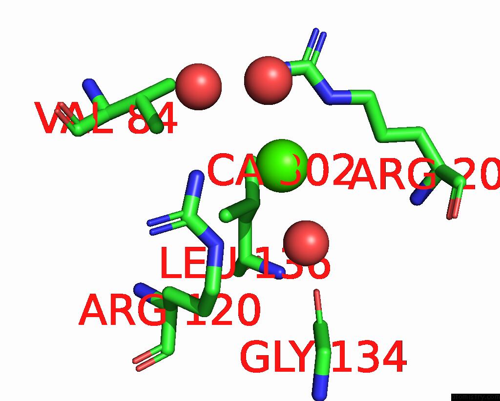
Mono view
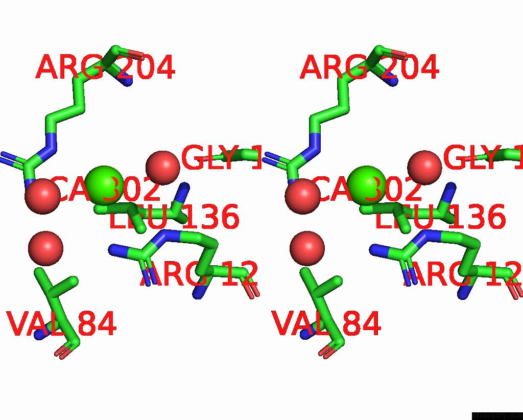
Stereo pair view
Mono view
Stereo pair view
A full contact list of Calcium with other atoms in the Ca binding
site number 1 of Crystal Structure of C-Terminal Domain of Pseudomonas Aeruginosa Lexa G91D Mutant within 5.0Å range:
|
Calcium binding site 2 out of 2 in 8b0v
Go back to
Calcium binding site 2 out
of 2 in the Crystal Structure of C-Terminal Domain of Pseudomonas Aeruginosa Lexa G91D Mutant
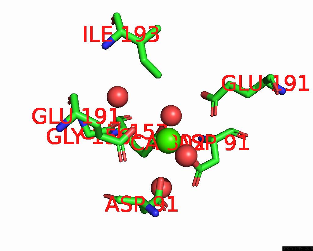
Mono view
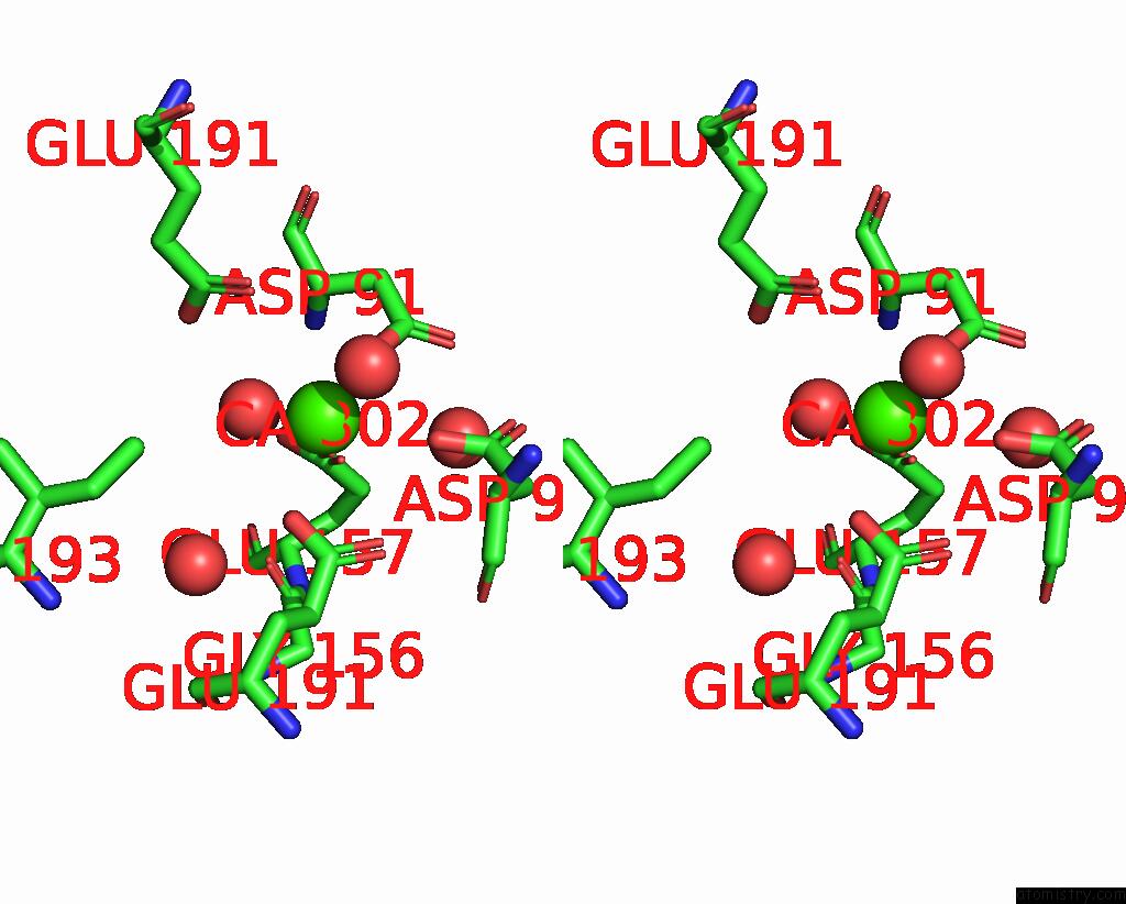
Stereo pair view
Mono view
Stereo pair view
A full contact list of Calcium with other atoms in the Ca binding
site number 2 of Crystal Structure of C-Terminal Domain of Pseudomonas Aeruginosa Lexa G91D Mutant within 5.0Å range:
|
Reference:
F.Vascon,
S.De Felice,
M.Gasparotto,
S.T.Huber,
C.Catalano,
M.Chinellato,
A.Grinzato,
F.Filippini,
L.Maso,
A.Jakobi,
L.Cendron.
Structural Investigations on the Sos Response in Pseudomonas Aeruginosa To Be Published.
Page generated: Thu Jul 10 03:18:08 2025
Last articles
Cd in 2PZICd in 2P64
Cd in 2OTJ
Cd in 2OTL
Cd in 2OZQ
Cd in 2NNE
Cd in 2MRB
Cd in 2MRT
Cd in 2NND
Cd in 2MPA