Calcium »
PDB 8ax0-8bad »
8b53 »
Calcium in PDB 8b53: Structure of Porcine Pancreatic Elastase Bound to A Fragment of A 4- Azaindole Inhibitor
Enzymatic activity of Structure of Porcine Pancreatic Elastase Bound to A Fragment of A 4- Azaindole Inhibitor
All present enzymatic activity of Structure of Porcine Pancreatic Elastase Bound to A Fragment of A 4- Azaindole Inhibitor:
3.4.21.36;
3.4.21.36;
Protein crystallography data
The structure of Structure of Porcine Pancreatic Elastase Bound to A Fragment of A 4- Azaindole Inhibitor, PDB code: 8b53
was solved by
M.Ferraroni,
A.Gerace,
with X-Ray Crystallography technique. A brief refinement statistics is given in the table below:
| Resolution Low / High (Å) | 22.25 / 1.25 |
| Space group | P 21 21 21 |
| Cell size a, b, c (Å), α, β, γ (°) | 50.019, 57.91, 74.519, 90, 90, 90 |
| R / Rfree (%) | 11.2 / 13.5 |
Calcium Binding Sites:
The binding sites of Calcium atom in the Structure of Porcine Pancreatic Elastase Bound to A Fragment of A 4- Azaindole Inhibitor
(pdb code 8b53). This binding sites where shown within
5.0 Angstroms radius around Calcium atom.
In total only one binding site of Calcium was determined in the Structure of Porcine Pancreatic Elastase Bound to A Fragment of A 4- Azaindole Inhibitor, PDB code: 8b53:
In total only one binding site of Calcium was determined in the Structure of Porcine Pancreatic Elastase Bound to A Fragment of A 4- Azaindole Inhibitor, PDB code: 8b53:
Calcium binding site 1 out of 1 in 8b53
Go back to
Calcium binding site 1 out
of 1 in the Structure of Porcine Pancreatic Elastase Bound to A Fragment of A 4- Azaindole Inhibitor
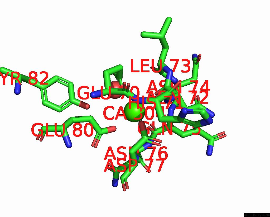
Mono view
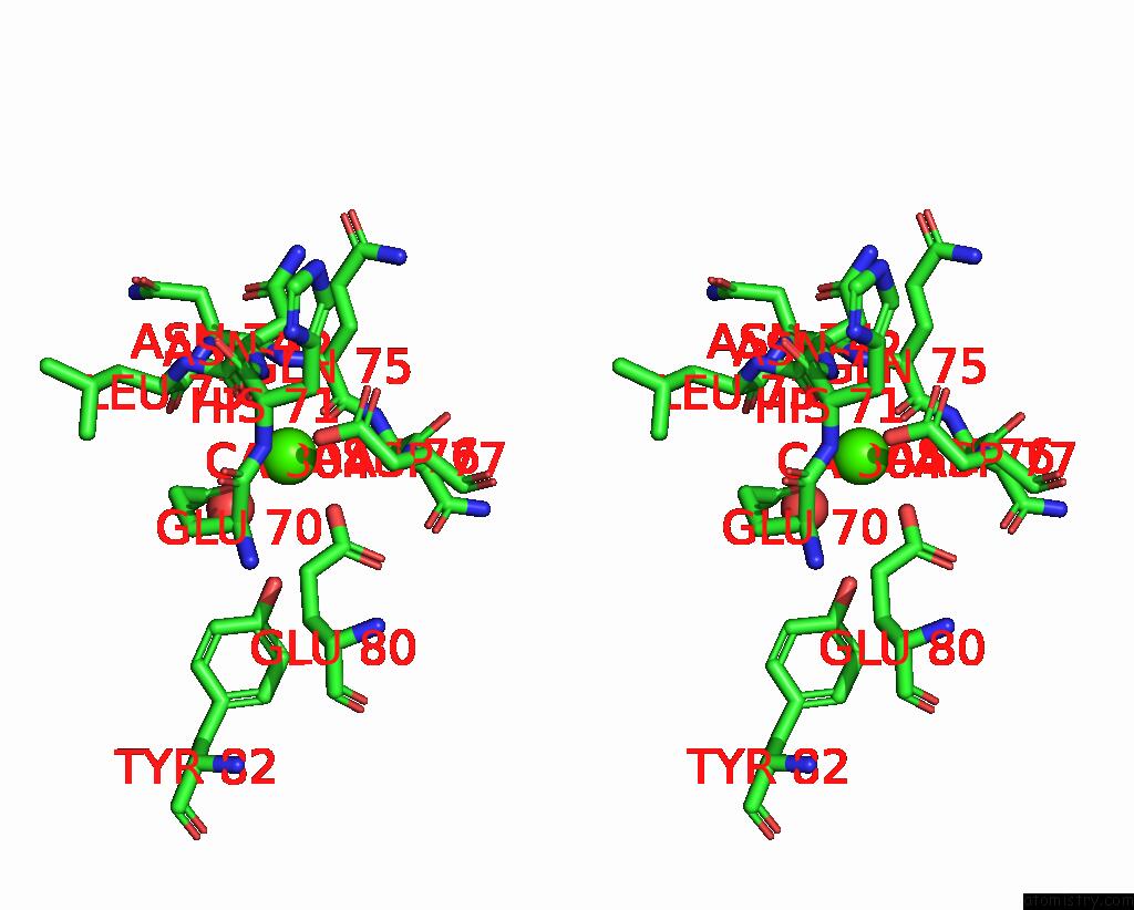
Stereo pair view
Mono view
Stereo pair view
A full contact list of Calcium with other atoms in the Ca binding
site number 1 of Structure of Porcine Pancreatic Elastase Bound to A Fragment of A 4- Azaindole Inhibitor within 5.0Å range:
|
Reference:
A.Gerace,
V.Masini,
L.Crocetti,
M.P.Giovannoni,
M.Ferraroni.
X-Ray Structural Study of Human Neutrophil Elastase Inhibition with A Series of Azaindoles, Azaindazoles and Isoxazolones J.Mol.Struct. V.1274 34595 2023.
ISSN: ISSN 0022-2860
DOI: 10.1016/J.MOLSTRUC.2022.134595
Page generated: Thu Jul 10 03:20:38 2025
ISSN: ISSN 0022-2860
DOI: 10.1016/J.MOLSTRUC.2022.134595
Last articles
Cd in 3ZI7Cd in 3ZG5
Cd in 3ZFZ
Cd in 3ZG0
Cd in 3WZO
Cd in 3ZET
Cd in 3WNK
Cd in 3X1Y
Cd in 3X1X
Cd in 3X1W