Calcium in PDB 8b8j: Cryo-Em Structure of CA2+-Bound MTMEM16F F518H Mutant in Digitonin
Calcium Binding Sites:
The binding sites of Calcium atom in the Cryo-Em Structure of CA2+-Bound MTMEM16F F518H Mutant in Digitonin
(pdb code 8b8j). This binding sites where shown within
5.0 Angstroms radius around Calcium atom.
In total 6 binding sites of Calcium where determined in the
Cryo-Em Structure of CA2+-Bound MTMEM16F F518H Mutant in Digitonin, PDB code: 8b8j:
Jump to Calcium binding site number:
1;
2;
3;
4;
5;
6;
Calcium binding site 1 out
of 6 in 8b8j
Go back to
Calcium Binding Sites List in 8b8j
Calcium binding site 1 out
of 6 in the Cryo-Em Structure of CA2+-Bound MTMEM16F F518H Mutant in Digitonin
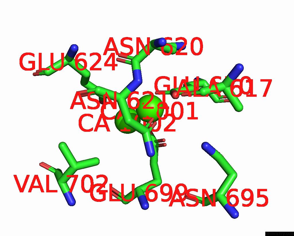
Mono view
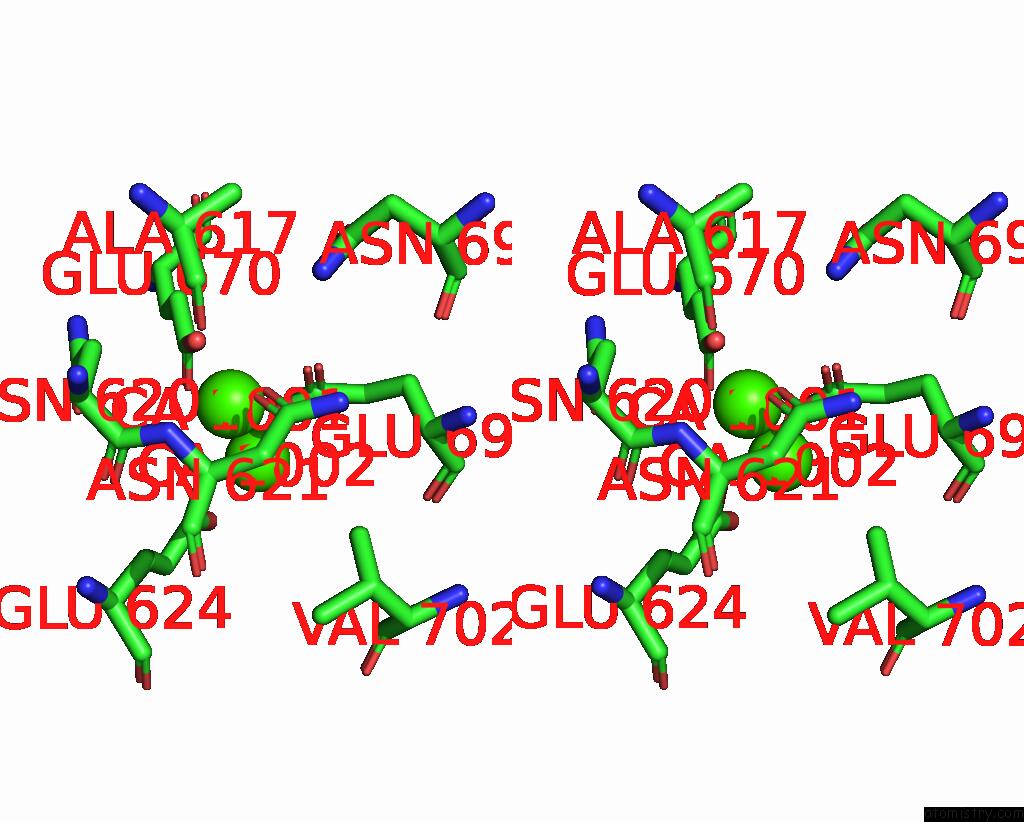
Stereo pair view
|
A full contact list of Calcium with other atoms in the Ca binding
site number 1 of Cryo-Em Structure of CA2+-Bound MTMEM16F F518H Mutant in Digitonin within 5.0Å range:
probe
|
atom
|
residue
|
distance (Å)
|
B
|
Occ
|
A:Ca1001
b:133.0
occ:1.00
|
OD1
|
A:ASN621
|
2.3
|
116.8
|
1.0
|
OE2
|
A:GLU670
|
2.3
|
108.2
|
1.0
|
OE2
|
A:GLU699
|
2.3
|
112.4
|
1.0
|
OE1
|
A:GLU699
|
2.4
|
106.5
|
1.0
|
OE1
|
A:GLU624
|
2.5
|
107.1
|
1.0
|
CD
|
A:GLU699
|
2.7
|
106.8
|
1.0
|
CD
|
A:GLU670
|
3.2
|
110.8
|
1.0
|
OE1
|
A:GLU670
|
3.3
|
107.5
|
1.0
|
CG
|
A:ASN621
|
3.4
|
118.9
|
1.0
|
CD
|
A:GLU624
|
3.8
|
113.2
|
1.0
|
CA
|
A:ASN621
|
3.9
|
111.0
|
1.0
|
N
|
A:ASN621
|
4.0
|
112.3
|
1.0
|
CA
|
A:CA1002
|
4.0
|
88.0
|
1.0
|
OD1
|
A:ASN620
|
4.1
|
124.5
|
1.0
|
CG
|
A:ASN620
|
4.1
|
125.3
|
1.0
|
CG
|
A:GLU699
|
4.2
|
96.0
|
1.0
|
C
|
A:ASN620
|
4.2
|
116.4
|
1.0
|
CB
|
A:ASN621
|
4.2
|
115.7
|
1.0
|
ND2
|
A:ASN621
|
4.3
|
120.9
|
1.0
|
O
|
A:ASN620
|
4.4
|
122.2
|
1.0
|
OE2
|
A:GLU624
|
4.4
|
115.8
|
1.0
|
CB
|
A:ASN620
|
4.5
|
120.1
|
1.0
|
ND2
|
A:ASN620
|
4.6
|
121.4
|
1.0
|
CG
|
A:GLU670
|
4.6
|
106.2
|
1.0
|
CB
|
A:GLU624
|
4.7
|
115.1
|
1.0
|
ND2
|
A:ASN695
|
4.8
|
99.9
|
1.0
|
CG
|
A:GLU624
|
4.8
|
119.1
|
1.0
|
O
|
A:ALA617
|
4.9
|
118.9
|
1.0
|
CG1
|
A:VAL702
|
4.9
|
102.8
|
1.0
|
CB
|
A:GLU699
|
4.9
|
99.2
|
1.0
|
|
Calcium binding site 2 out
of 6 in 8b8j
Go back to
Calcium Binding Sites List in 8b8j
Calcium binding site 2 out
of 6 in the Cryo-Em Structure of CA2+-Bound MTMEM16F F518H Mutant in Digitonin
Mono view
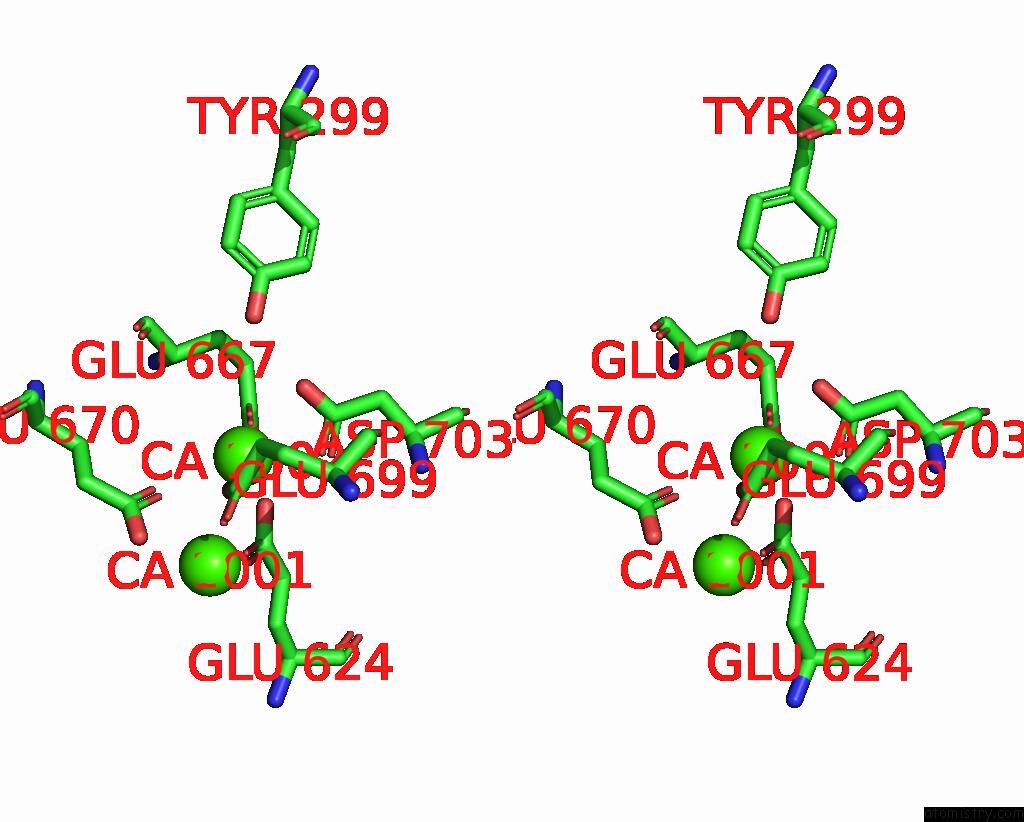
Stereo pair view
|
A full contact list of Calcium with other atoms in the Ca binding
site number 2 of Cryo-Em Structure of CA2+-Bound MTMEM16F F518H Mutant in Digitonin within 5.0Å range:
probe
|
atom
|
residue
|
distance (Å)
|
B
|
Occ
|
A:Ca1002
b:88.0
occ:1.00
|
OE1
|
A:GLU667
|
2.3
|
121.6
|
1.0
|
OE2
|
A:GLU624
|
2.3
|
115.8
|
1.0
|
OE2
|
A:GLU699
|
2.3
|
112.4
|
1.0
|
OD1
|
A:ASP703
|
2.5
|
109.0
|
1.0
|
OD2
|
A:ASP703
|
2.8
|
105.2
|
1.0
|
OE1
|
A:GLU670
|
2.9
|
107.5
|
1.0
|
CG
|
A:ASP703
|
3.1
|
104.0
|
1.0
|
CD
|
A:GLU624
|
3.1
|
113.2
|
1.0
|
OE1
|
A:GLU624
|
3.1
|
107.1
|
1.0
|
CD
|
A:GLU699
|
3.4
|
106.8
|
1.0
|
CD
|
A:GLU667
|
3.5
|
121.9
|
1.0
|
CG
|
A:GLU699
|
3.8
|
96.0
|
1.0
|
CD
|
A:GLU670
|
3.9
|
110.8
|
1.0
|
CA
|
A:CA1001
|
4.0
|
133.0
|
1.0
|
OE2
|
A:GLU667
|
4.2
|
125.0
|
1.0
|
OH
|
A:TYR299
|
4.3
|
100.0
|
1.0
|
OE2
|
A:GLU670
|
4.4
|
108.2
|
1.0
|
OE1
|
A:GLU699
|
4.5
|
106.5
|
1.0
|
CG
|
A:GLU624
|
4.5
|
119.1
|
1.0
|
CA
|
A:GLU667
|
4.6
|
107.4
|
1.0
|
CB
|
A:ASP703
|
4.6
|
92.7
|
1.0
|
CB
|
A:GLU667
|
4.6
|
110.3
|
1.0
|
CG
|
A:GLU667
|
4.6
|
114.6
|
1.0
|
|
Calcium binding site 3 out
of 6 in 8b8j
Go back to
Calcium Binding Sites List in 8b8j
Calcium binding site 3 out
of 6 in the Cryo-Em Structure of CA2+-Bound MTMEM16F F518H Mutant in Digitonin
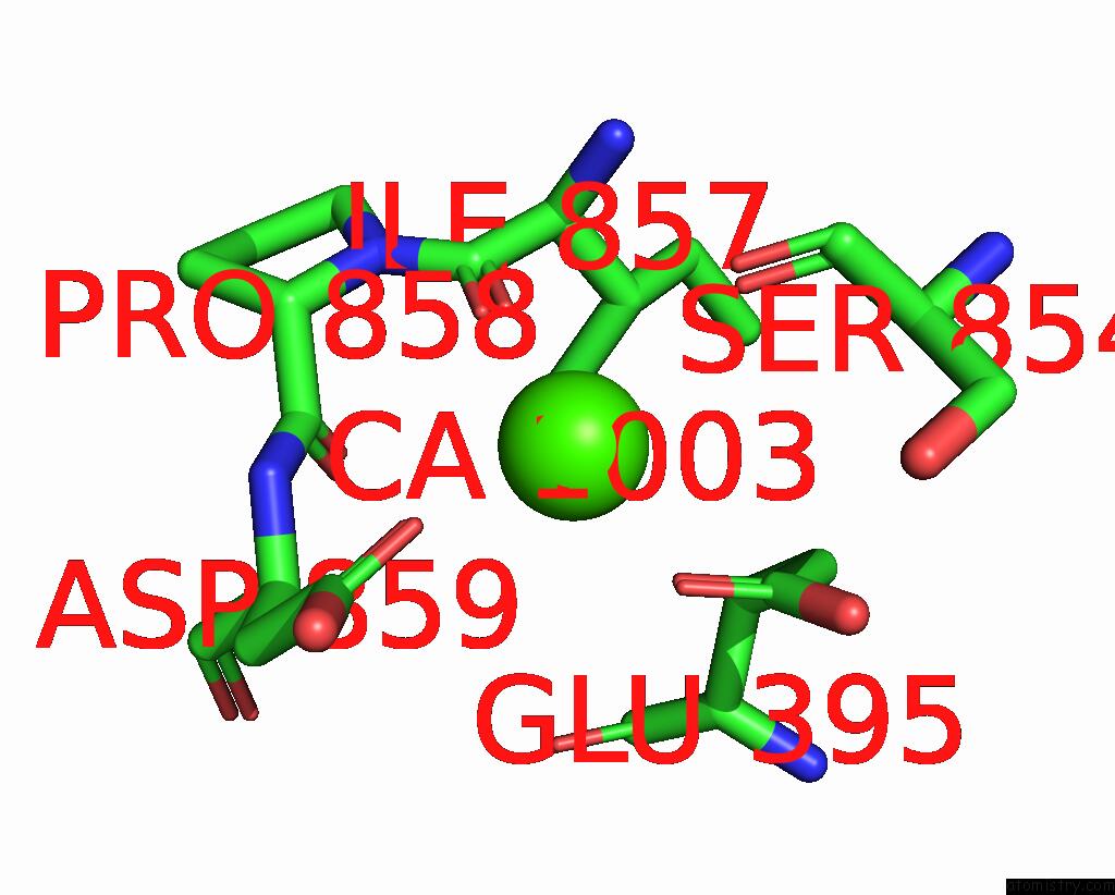
Mono view
Stereo pair view
|
A full contact list of Calcium with other atoms in the Ca binding
site number 3 of Cryo-Em Structure of CA2+-Bound MTMEM16F F518H Mutant in Digitonin within 5.0Å range:
probe
|
atom
|
residue
|
distance (Å)
|
B
|
Occ
|
A:Ca1003
b:153.6
occ:1.00
|
O
|
A:ILE857
|
2.3
|
127.4
|
1.0
|
OD1
|
A:ASP859
|
2.4
|
154.4
|
1.0
|
OE1
|
A:GLU395
|
2.7
|
134.3
|
1.0
|
O
|
A:SER854
|
2.9
|
122.2
|
1.0
|
C
|
A:ILE857
|
3.5
|
127.2
|
1.0
|
CG
|
A:ASP859
|
3.6
|
152.8
|
1.0
|
CD
|
A:GLU395
|
3.6
|
133.8
|
1.0
|
N
|
A:ASP859
|
3.9
|
146.5
|
1.0
|
C
|
A:SER854
|
4.0
|
123.8
|
1.0
|
C
|
A:PRO858
|
4.0
|
143.7
|
1.0
|
OE2
|
A:GLU395
|
4.0
|
130.9
|
1.0
|
CA
|
A:ASP859
|
4.2
|
147.8
|
1.0
|
CB
|
A:ILE857
|
4.3
|
124.5
|
1.0
|
OG
|
A:SER854
|
4.3
|
120.2
|
1.0
|
CA
|
A:ILE857
|
4.3
|
123.7
|
1.0
|
CG2
|
A:ILE857
|
4.4
|
124.5
|
1.0
|
O
|
A:PRO858
|
4.4
|
146.8
|
1.0
|
OD2
|
A:ASP859
|
4.4
|
151.5
|
1.0
|
N
|
A:PRO858
|
4.4
|
146.4
|
1.0
|
CA
|
A:PRO858
|
4.5
|
145.2
|
1.0
|
CB
|
A:ASP859
|
4.5
|
150.4
|
1.0
|
CA
|
A:SER854
|
4.5
|
118.3
|
1.0
|
N
|
A:ILE857
|
4.5
|
123.6
|
1.0
|
CG
|
A:GLU395
|
4.7
|
131.7
|
1.0
|
|
Calcium binding site 4 out
of 6 in 8b8j
Go back to
Calcium Binding Sites List in 8b8j
Calcium binding site 4 out
of 6 in the Cryo-Em Structure of CA2+-Bound MTMEM16F F518H Mutant in Digitonin
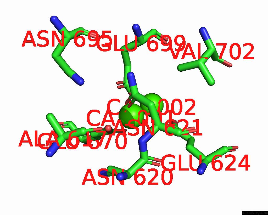
Mono view
Stereo pair view
|
A full contact list of Calcium with other atoms in the Ca binding
site number 4 of Cryo-Em Structure of CA2+-Bound MTMEM16F F518H Mutant in Digitonin within 5.0Å range:
probe
|
atom
|
residue
|
distance (Å)
|
B
|
Occ
|
B:Ca1001
b:133.0
occ:1.00
|
OD1
|
B:ASN621
|
2.3
|
116.8
|
1.0
|
OE2
|
B:GLU670
|
2.3
|
108.2
|
1.0
|
OE2
|
B:GLU699
|
2.3
|
112.4
|
1.0
|
OE1
|
B:GLU699
|
2.4
|
106.5
|
1.0
|
OE1
|
B:GLU624
|
2.5
|
107.1
|
1.0
|
CD
|
B:GLU699
|
2.7
|
106.8
|
1.0
|
CD
|
B:GLU670
|
3.2
|
110.8
|
1.0
|
OE1
|
B:GLU670
|
3.3
|
107.5
|
1.0
|
CG
|
B:ASN621
|
3.4
|
118.9
|
1.0
|
CD
|
B:GLU624
|
3.8
|
113.2
|
1.0
|
CA
|
B:ASN621
|
3.9
|
111.0
|
1.0
|
N
|
B:ASN621
|
4.0
|
112.3
|
1.0
|
CA
|
B:CA1002
|
4.0
|
88.0
|
1.0
|
OD1
|
B:ASN620
|
4.1
|
124.5
|
1.0
|
CG
|
B:ASN620
|
4.1
|
125.3
|
1.0
|
CG
|
B:GLU699
|
4.2
|
96.0
|
1.0
|
C
|
B:ASN620
|
4.2
|
116.4
|
1.0
|
CB
|
B:ASN621
|
4.2
|
115.7
|
1.0
|
ND2
|
B:ASN621
|
4.3
|
120.9
|
1.0
|
O
|
B:ASN620
|
4.4
|
122.2
|
1.0
|
OE2
|
B:GLU624
|
4.4
|
115.8
|
1.0
|
CB
|
B:ASN620
|
4.5
|
120.1
|
1.0
|
ND2
|
B:ASN620
|
4.6
|
121.4
|
1.0
|
CG
|
B:GLU670
|
4.6
|
106.2
|
1.0
|
CB
|
B:GLU624
|
4.7
|
115.1
|
1.0
|
ND2
|
B:ASN695
|
4.8
|
99.9
|
1.0
|
CG
|
B:GLU624
|
4.8
|
119.1
|
1.0
|
O
|
B:ALA617
|
4.9
|
118.9
|
1.0
|
CG1
|
B:VAL702
|
4.9
|
102.8
|
1.0
|
CB
|
B:GLU699
|
4.9
|
99.2
|
1.0
|
|
Calcium binding site 5 out
of 6 in 8b8j
Go back to
Calcium Binding Sites List in 8b8j
Calcium binding site 5 out
of 6 in the Cryo-Em Structure of CA2+-Bound MTMEM16F F518H Mutant in Digitonin
Mono view
Stereo pair view
|
A full contact list of Calcium with other atoms in the Ca binding
site number 5 of Cryo-Em Structure of CA2+-Bound MTMEM16F F518H Mutant in Digitonin within 5.0Å range:
probe
|
atom
|
residue
|
distance (Å)
|
B
|
Occ
|
B:Ca1002
b:88.0
occ:1.00
|
OE1
|
B:GLU667
|
2.3
|
121.6
|
1.0
|
OE2
|
B:GLU624
|
2.3
|
115.8
|
1.0
|
OE2
|
B:GLU699
|
2.3
|
112.4
|
1.0
|
OD1
|
B:ASP703
|
2.5
|
109.0
|
1.0
|
OD2
|
B:ASP703
|
2.8
|
105.2
|
1.0
|
OE1
|
B:GLU670
|
2.9
|
107.5
|
1.0
|
CG
|
B:ASP703
|
3.1
|
104.0
|
1.0
|
CD
|
B:GLU624
|
3.1
|
113.2
|
1.0
|
OE1
|
B:GLU624
|
3.1
|
107.1
|
1.0
|
CD
|
B:GLU699
|
3.4
|
106.8
|
1.0
|
CD
|
B:GLU667
|
3.5
|
121.9
|
1.0
|
CG
|
B:GLU699
|
3.8
|
96.0
|
1.0
|
CD
|
B:GLU670
|
3.9
|
110.8
|
1.0
|
CA
|
B:CA1001
|
4.0
|
133.0
|
1.0
|
OE2
|
B:GLU667
|
4.2
|
125.0
|
1.0
|
OH
|
B:TYR299
|
4.3
|
100.0
|
1.0
|
OE2
|
B:GLU670
|
4.4
|
108.2
|
1.0
|
OE1
|
B:GLU699
|
4.5
|
106.5
|
1.0
|
CG
|
B:GLU624
|
4.5
|
119.1
|
1.0
|
CA
|
B:GLU667
|
4.6
|
107.4
|
1.0
|
CB
|
B:ASP703
|
4.6
|
92.7
|
1.0
|
CB
|
B:GLU667
|
4.6
|
110.3
|
1.0
|
CG
|
B:GLU667
|
4.6
|
114.6
|
1.0
|
|
Calcium binding site 6 out
of 6 in 8b8j
Go back to
Calcium Binding Sites List in 8b8j
Calcium binding site 6 out
of 6 in the Cryo-Em Structure of CA2+-Bound MTMEM16F F518H Mutant in Digitonin
Mono view
Stereo pair view
|
A full contact list of Calcium with other atoms in the Ca binding
site number 6 of Cryo-Em Structure of CA2+-Bound MTMEM16F F518H Mutant in Digitonin within 5.0Å range:
probe
|
atom
|
residue
|
distance (Å)
|
B
|
Occ
|
B:Ca1003
b:153.6
occ:1.00
|
O
|
B:ILE857
|
2.3
|
127.4
|
1.0
|
OD1
|
B:ASP859
|
2.4
|
154.4
|
1.0
|
OE1
|
B:GLU395
|
2.7
|
134.3
|
1.0
|
O
|
B:SER854
|
2.9
|
122.2
|
1.0
|
C
|
B:ILE857
|
3.5
|
127.2
|
1.0
|
CG
|
B:ASP859
|
3.6
|
152.8
|
1.0
|
CD
|
B:GLU395
|
3.6
|
133.8
|
1.0
|
N
|
B:ASP859
|
3.9
|
146.5
|
1.0
|
C
|
B:SER854
|
4.0
|
123.8
|
1.0
|
C
|
B:PRO858
|
4.0
|
143.7
|
1.0
|
OE2
|
B:GLU395
|
4.0
|
130.9
|
1.0
|
CA
|
B:ASP859
|
4.2
|
147.8
|
1.0
|
CB
|
B:ILE857
|
4.3
|
124.5
|
1.0
|
OG
|
B:SER854
|
4.3
|
120.2
|
1.0
|
CA
|
B:ILE857
|
4.3
|
123.7
|
1.0
|
CG2
|
B:ILE857
|
4.4
|
124.5
|
1.0
|
O
|
B:PRO858
|
4.4
|
146.8
|
1.0
|
OD2
|
B:ASP859
|
4.4
|
151.5
|
1.0
|
N
|
B:PRO858
|
4.4
|
146.4
|
1.0
|
CA
|
B:PRO858
|
4.5
|
145.2
|
1.0
|
CB
|
B:ASP859
|
4.5
|
150.4
|
1.0
|
CA
|
B:SER854
|
4.5
|
118.3
|
1.0
|
N
|
B:ILE857
|
4.5
|
123.6
|
1.0
|
CG
|
B:GLU395
|
4.7
|
131.7
|
1.0
|
|
Reference:
M.Arndt,
C.Alvadia,
M.S.Straub,
V.Clerico Mosina,
C.Paulino,
R.Dutzler.
Structural Basis For the Activation of the Lipid Scramblase TMEM16F. Nat Commun V. 13 6692 2022.
ISSN: ESSN 2041-1723
PubMed: 36335104
DOI: 10.1038/S41467-022-34497-X
Page generated: Fri Jul 19 07:13:35 2024
|