Calcium »
PDB 8ban-8bsc »
8bay »
Calcium in PDB 8bay: Crystal Structure of IDH1 Variant R132C S280F in Complex with Nadph, CA2+ and 3-Butyl-2-Oxoglutarate
Enzymatic activity of Crystal Structure of IDH1 Variant R132C S280F in Complex with Nadph, CA2+ and 3-Butyl-2-Oxoglutarate
All present enzymatic activity of Crystal Structure of IDH1 Variant R132C S280F in Complex with Nadph, CA2+ and 3-Butyl-2-Oxoglutarate:
1.1.1.42;
1.1.1.42;
Protein crystallography data
The structure of Crystal Structure of IDH1 Variant R132C S280F in Complex with Nadph, CA2+ and 3-Butyl-2-Oxoglutarate, PDB code: 8bay
was solved by
P.Rabe,
C.J.Schofield,
R.Reinbold,
L.Brewitz,
with X-Ray Crystallography technique. A brief refinement statistics is given in the table below:
| Resolution Low / High (Å) | 46.81 / 2.35 |
| Space group | C 2 2 21 |
| Cell size a, b, c (Å), α, β, γ (°) | 99.54, 275.78, 116.61, 90, 90, 90 |
| R / Rfree (%) | 20.1 / 22.3 |
Calcium Binding Sites:
The binding sites of Calcium atom in the Crystal Structure of IDH1 Variant R132C S280F in Complex with Nadph, CA2+ and 3-Butyl-2-Oxoglutarate
(pdb code 8bay). This binding sites where shown within
5.0 Angstroms radius around Calcium atom.
In total 3 binding sites of Calcium where determined in the Crystal Structure of IDH1 Variant R132C S280F in Complex with Nadph, CA2+ and 3-Butyl-2-Oxoglutarate, PDB code: 8bay:
Jump to Calcium binding site number: 1; 2; 3;
In total 3 binding sites of Calcium where determined in the Crystal Structure of IDH1 Variant R132C S280F in Complex with Nadph, CA2+ and 3-Butyl-2-Oxoglutarate, PDB code: 8bay:
Jump to Calcium binding site number: 1; 2; 3;
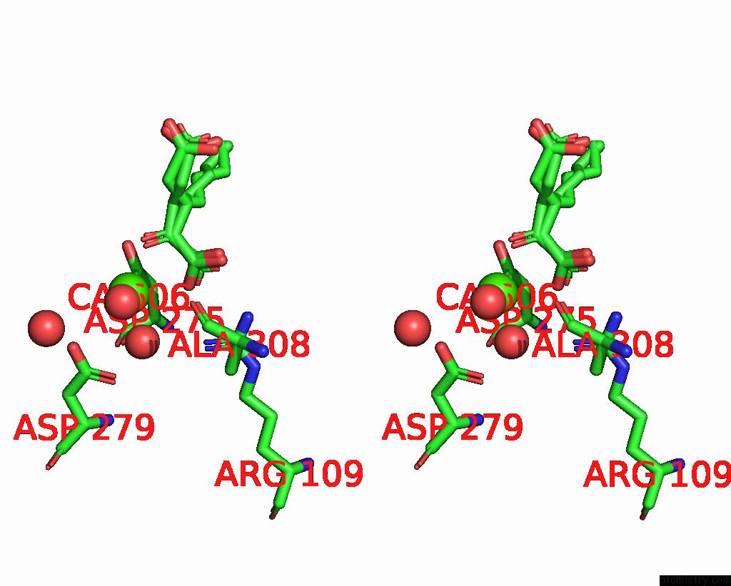
Calcium binding site 1 out of 3 in 8bay
Go back to
Calcium binding site 1 out
of 3 in the Crystal Structure of IDH1 Variant R132C S280F in Complex with Nadph, CA2+ and 3-Butyl-2-Oxoglutarate
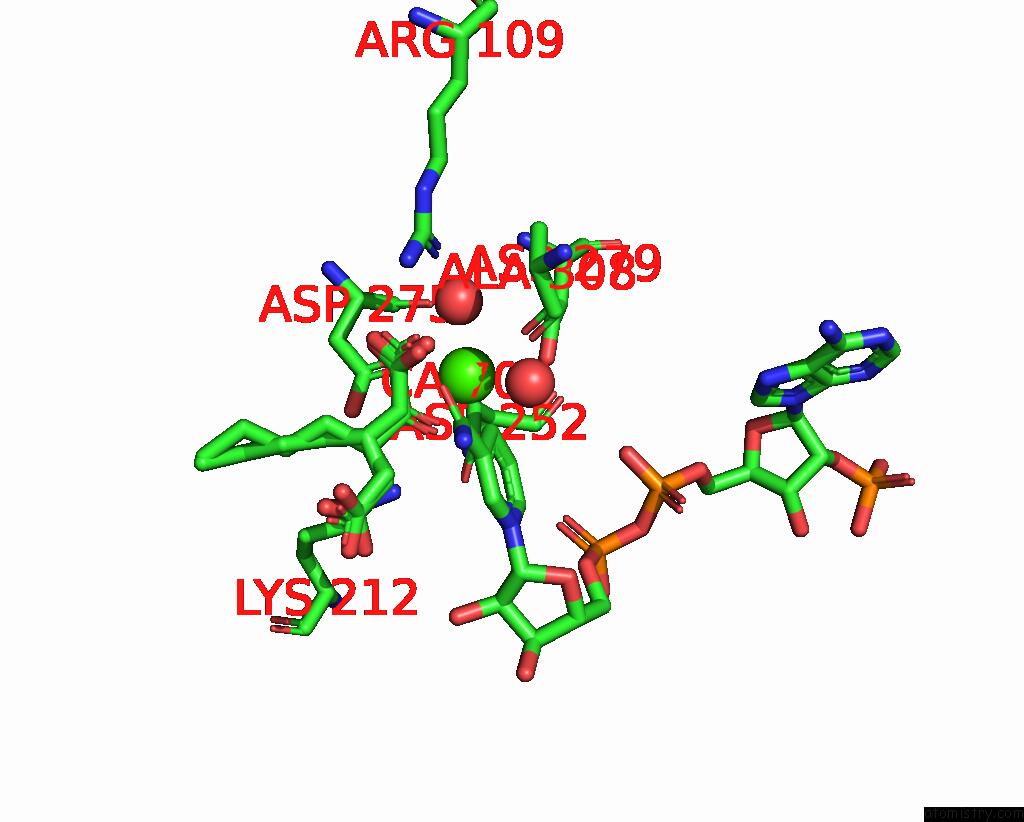
Mono view

Stereo pair view
Mono view
Stereo pair view
A full contact list of Calcium with other atoms in the Ca binding
site number 1 of Crystal Structure of IDH1 Variant R132C S280F in Complex with Nadph, CA2+ and 3-Butyl-2-Oxoglutarate within 5.0Å range:
|
Calcium binding site 2 out of 3 in 8bay
Go back to
Calcium binding site 2 out
of 3 in the Crystal Structure of IDH1 Variant R132C S280F in Complex with Nadph, CA2+ and 3-Butyl-2-Oxoglutarate
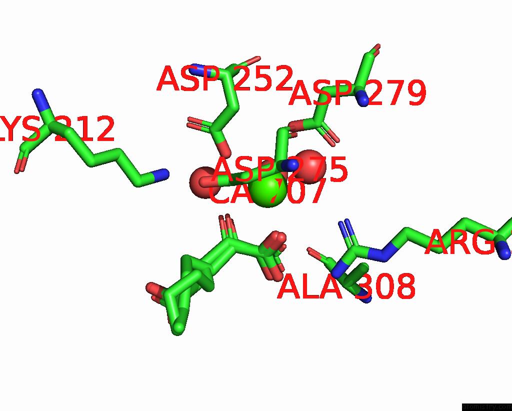
Mono view
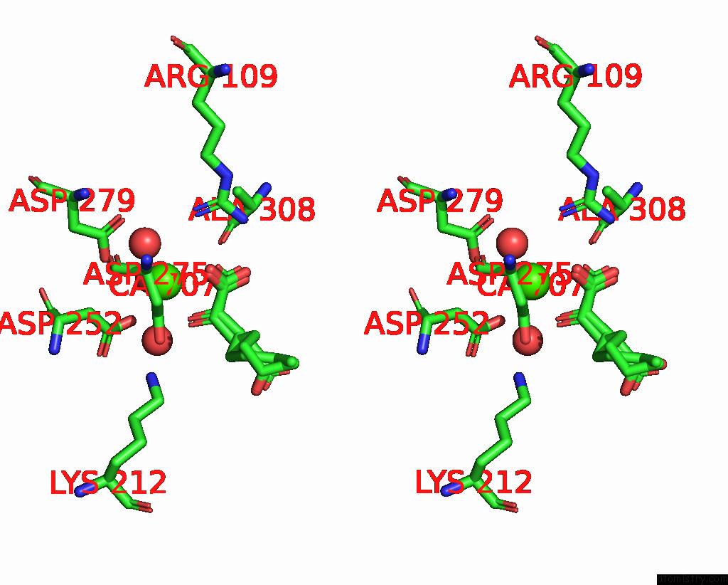
Stereo pair view
Mono view
Stereo pair view
A full contact list of Calcium with other atoms in the Ca binding
site number 2 of Crystal Structure of IDH1 Variant R132C S280F in Complex with Nadph, CA2+ and 3-Butyl-2-Oxoglutarate within 5.0Å range:
|
Calcium binding site 3 out of 3 in 8bay
Go back to
Calcium binding site 3 out
of 3 in the Crystal Structure of IDH1 Variant R132C S280F in Complex with Nadph, CA2+ and 3-Butyl-2-Oxoglutarate
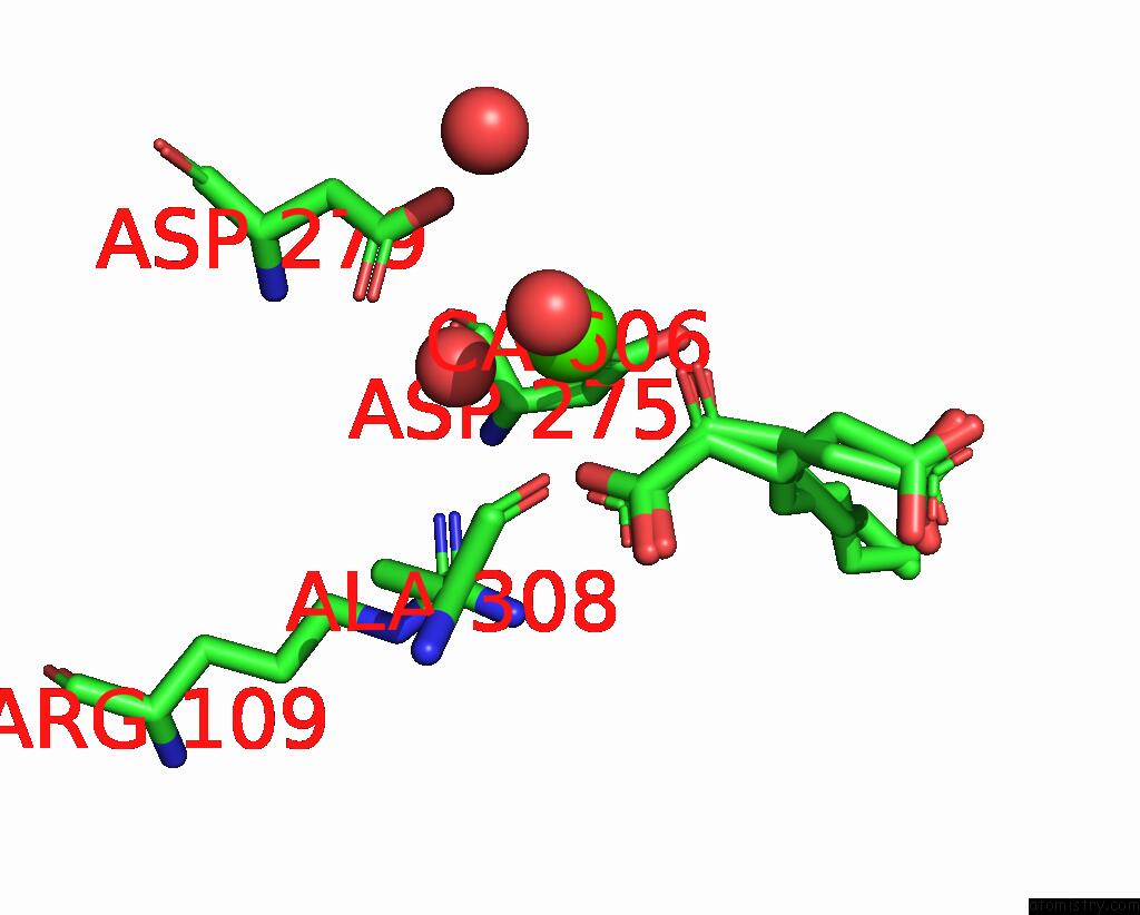
Mono view
Stereo pair view
Mono view
Stereo pair view
A full contact list of Calcium with other atoms in the Ca binding
site number 3 of Crystal Structure of IDH1 Variant R132C S280F in Complex with Nadph, CA2+ and 3-Butyl-2-Oxoglutarate within 5.0Å range:
|
Reference:
P.Rabe,
C.J.Schofield,
R.Reinbold,
L.Brewitz.
Crystal Structure of IDH1 Variant R132C S280F in Complex with Nadph, CA2+ and 3-Butyl-2-Oxoglutarate To Be Published.
Page generated: Thu Jul 10 03:25:23 2025
Last articles
Cl in 8CWJCl in 8CXJ
Cl in 8CXL
Cl in 8CWH
Cl in 8CX9
Cl in 8CWI
Cl in 8CX5
Cl in 8CWG
Cl in 8CWF
Cl in 8CWE