Calcium »
PDB 8ban-8bsc »
8bo8 »
Calcium in PDB 8bo8: X-Ray Structure of Human Glutamate Carboxypeptidase II (Gcpii) in Complex with An Inhibitor P17
Enzymatic activity of X-Ray Structure of Human Glutamate Carboxypeptidase II (Gcpii) in Complex with An Inhibitor P17
All present enzymatic activity of X-Ray Structure of Human Glutamate Carboxypeptidase II (Gcpii) in Complex with An Inhibitor P17:
3.4.17.21;
3.4.17.21;
Protein crystallography data
The structure of X-Ray Structure of Human Glutamate Carboxypeptidase II (Gcpii) in Complex with An Inhibitor P17, PDB code: 8bo8
was solved by
L.Motlova,
C.Barinka,
M.Benesova,
with X-Ray Crystallography technique. A brief refinement statistics is given in the table below:
| Resolution Low / High (Å) | 50.01 / 1.55 |
| Space group | I 2 2 2 |
| Cell size a, b, c (Å), α, β, γ (°) | 101.887, 130.286, 159.702, 90, 90, 90 |
| R / Rfree (%) | 20.2 / 22.6 |
Other elements in 8bo8:
The structure of X-Ray Structure of Human Glutamate Carboxypeptidase II (Gcpii) in Complex with An Inhibitor P17 also contains other interesting chemical elements:
| Zinc | (Zn) | 2 atoms |
| Sodium | (Na) | 1 atom |
| Chlorine | (Cl) | 1 atom |
Calcium Binding Sites:
The binding sites of Calcium atom in the X-Ray Structure of Human Glutamate Carboxypeptidase II (Gcpii) in Complex with An Inhibitor P17
(pdb code 8bo8). This binding sites where shown within
5.0 Angstroms radius around Calcium atom.
In total only one binding site of Calcium was determined in the X-Ray Structure of Human Glutamate Carboxypeptidase II (Gcpii) in Complex with An Inhibitor P17, PDB code: 8bo8:
In total only one binding site of Calcium was determined in the X-Ray Structure of Human Glutamate Carboxypeptidase II (Gcpii) in Complex with An Inhibitor P17, PDB code: 8bo8:
Calcium binding site 1 out of 1 in 8bo8
Go back to
Calcium binding site 1 out
of 1 in the X-Ray Structure of Human Glutamate Carboxypeptidase II (Gcpii) in Complex with An Inhibitor P17
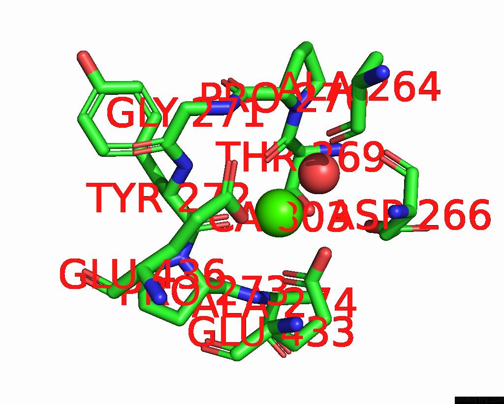
Mono view
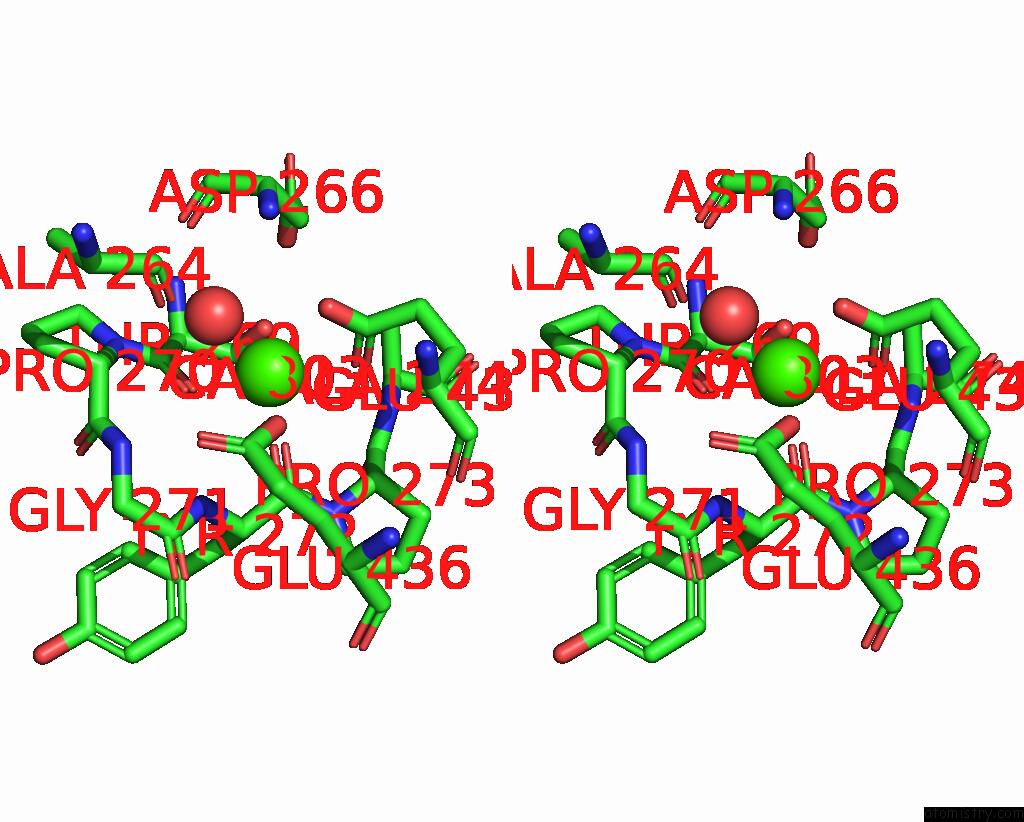
Stereo pair view
Mono view
Stereo pair view
A full contact list of Calcium with other atoms in the Ca binding
site number 1 of X-Ray Structure of Human Glutamate Carboxypeptidase II (Gcpii) in Complex with An Inhibitor P17 within 5.0Å range:
|
Reference:
L.Motlova,
C.Barinka,
M.Benesova.
X-Ray Structure of Human Glutamate Carboxypeptidase II (Gcpii) in Complex with An Inhibitor P17 To Be Published.
Page generated: Thu Jul 10 03:30:35 2025
Last articles
Cl in 5RZOCl in 5RZG
Cl in 5RYS
Cl in 5RYN
Cl in 5RY8
Cl in 5RY2
Cl in 5RY0
Cl in 5RXW
Cl in 5RXO
Cl in 5RXU