Calcium »
PDB 8bt4-8clf »
8cig »
Calcium in PDB 8cig: Crystal Structure of An 8-Repeat Consensus Tpr Superhelix in Tris Buffer with Calcium.
Protein crystallography data
The structure of Crystal Structure of An 8-Repeat Consensus Tpr Superhelix in Tris Buffer with Calcium., PDB code: 8cig
was solved by
M.Liutkus,
A.L.Rojas,
A.L.Cortajarena,
with X-Ray Crystallography technique. A brief refinement statistics is given in the table below:
| Resolution Low / High (Å) | 58.93 / 1.40 |
| Space group | P 32 2 1 |
| Cell size a, b, c (Å), α, β, γ (°) | 68.046, 68.046, 72.285, 90, 90, 120 |
| R / Rfree (%) | 16.9 / 19.2 |
Other elements in 8cig:
The structure of Crystal Structure of An 8-Repeat Consensus Tpr Superhelix in Tris Buffer with Calcium. also contains other interesting chemical elements:
| Chlorine | (Cl) | 2 atoms |
Calcium Binding Sites:
The binding sites of Calcium atom in the Crystal Structure of An 8-Repeat Consensus Tpr Superhelix in Tris Buffer with Calcium.
(pdb code 8cig). This binding sites where shown within
5.0 Angstroms radius around Calcium atom.
In total 6 binding sites of Calcium where determined in the Crystal Structure of An 8-Repeat Consensus Tpr Superhelix in Tris Buffer with Calcium., PDB code: 8cig:
Jump to Calcium binding site number: 1; 2; 3; 4; 5; 6;
In total 6 binding sites of Calcium where determined in the Crystal Structure of An 8-Repeat Consensus Tpr Superhelix in Tris Buffer with Calcium., PDB code: 8cig:
Jump to Calcium binding site number: 1; 2; 3; 4; 5; 6;
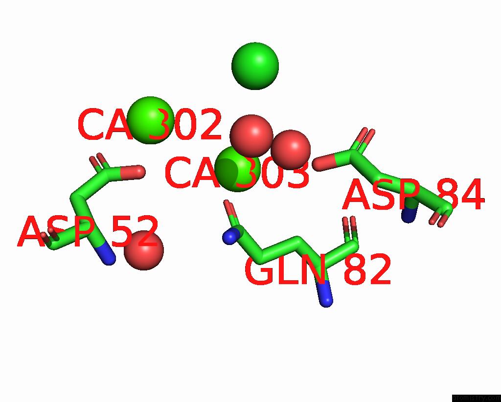
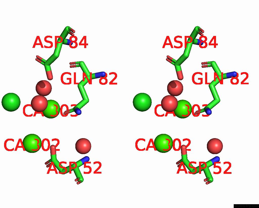
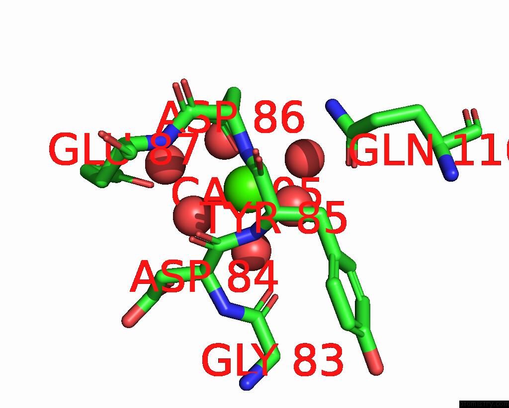
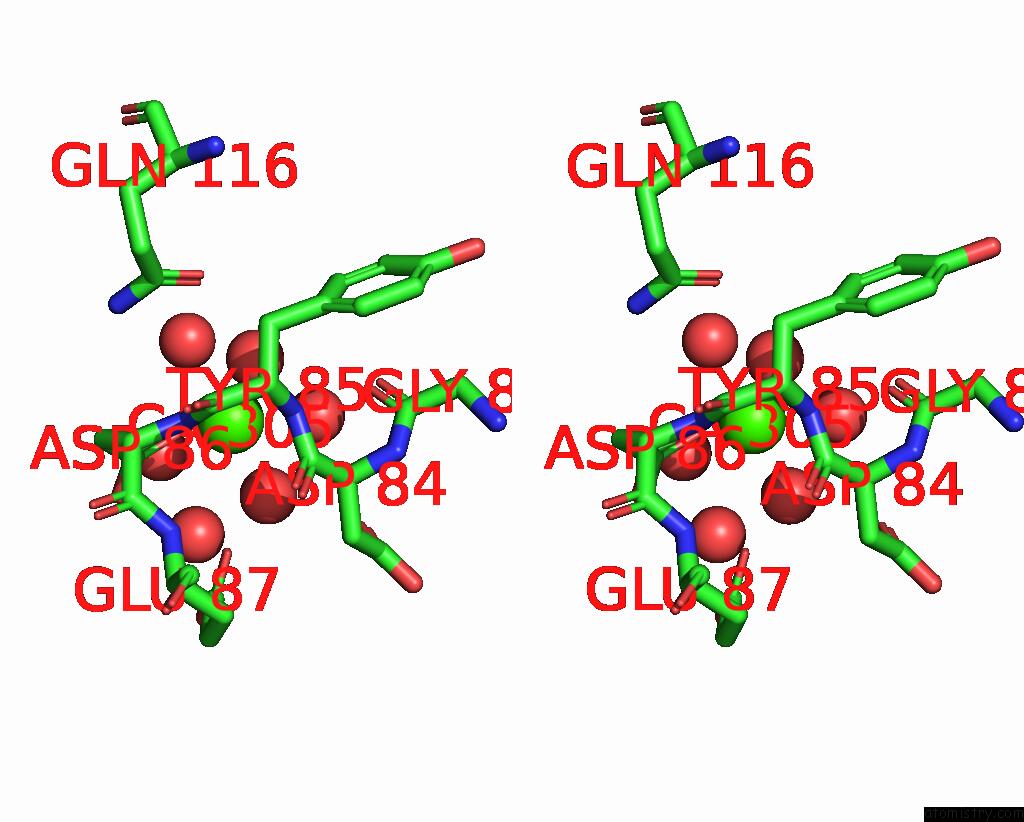
Calcium binding site 1 out of 6 in 8cig
Go back to
Calcium binding site 1 out
of 6 in the Crystal Structure of An 8-Repeat Consensus Tpr Superhelix in Tris Buffer with Calcium.
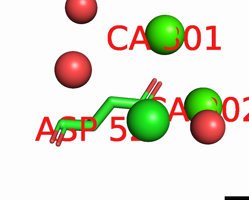
Mono view
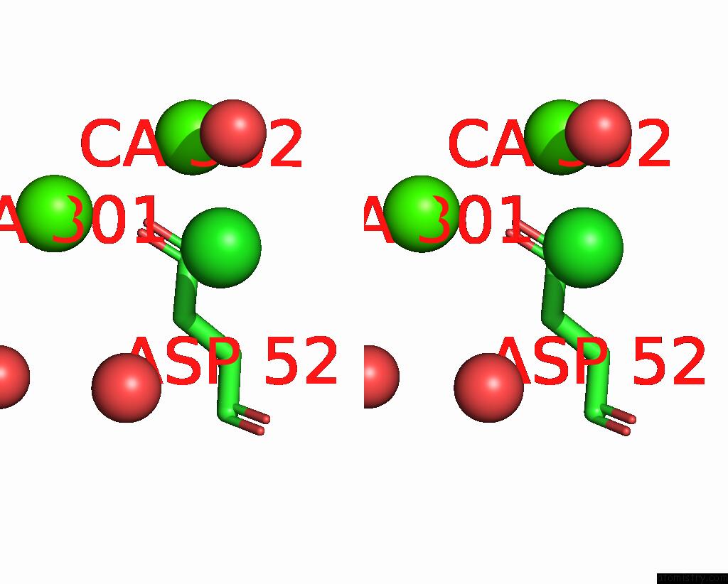
Stereo pair view
Mono view
Stereo pair view
A full contact list of Calcium with other atoms in the Ca binding
site number 1 of Crystal Structure of An 8-Repeat Consensus Tpr Superhelix in Tris Buffer with Calcium. within 5.0Å range:
|
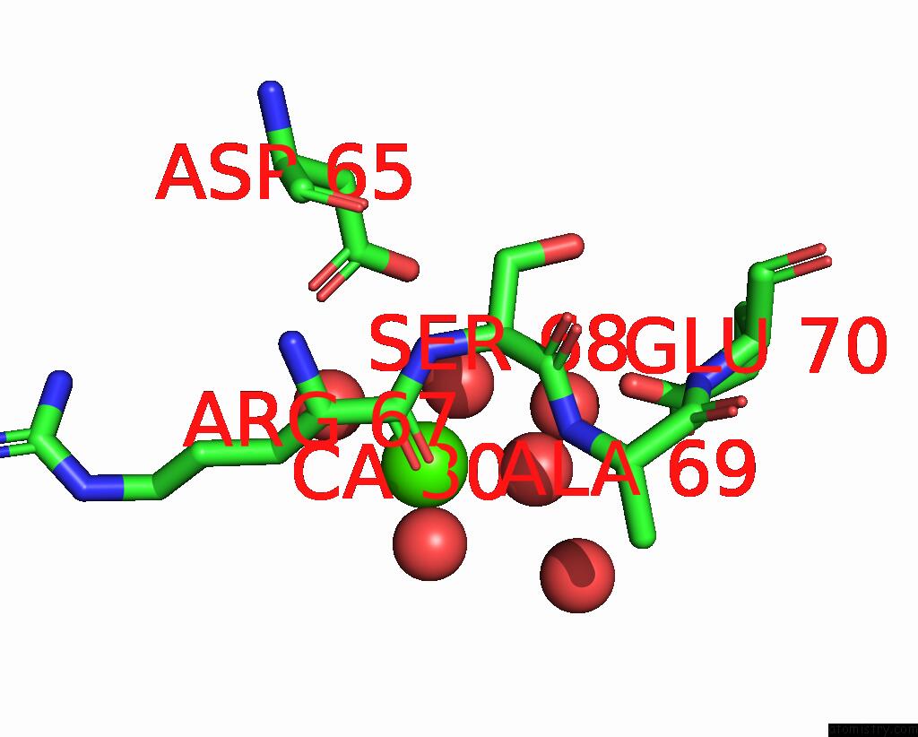
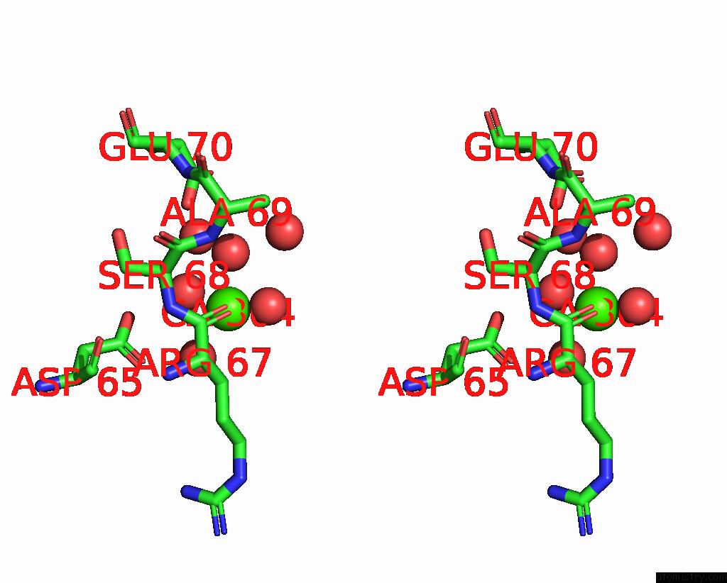
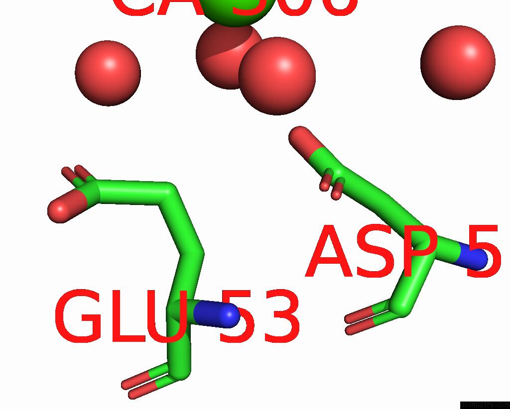
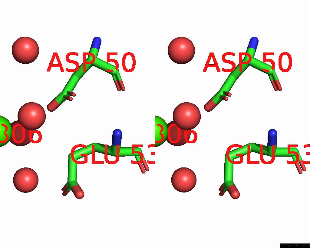
Calcium binding site 2 out of 6 in 8cig
Go back to
Calcium binding site 2 out
of 6 in the Crystal Structure of An 8-Repeat Consensus Tpr Superhelix in Tris Buffer with Calcium.
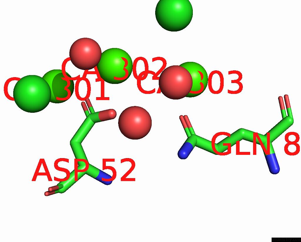
Mono view
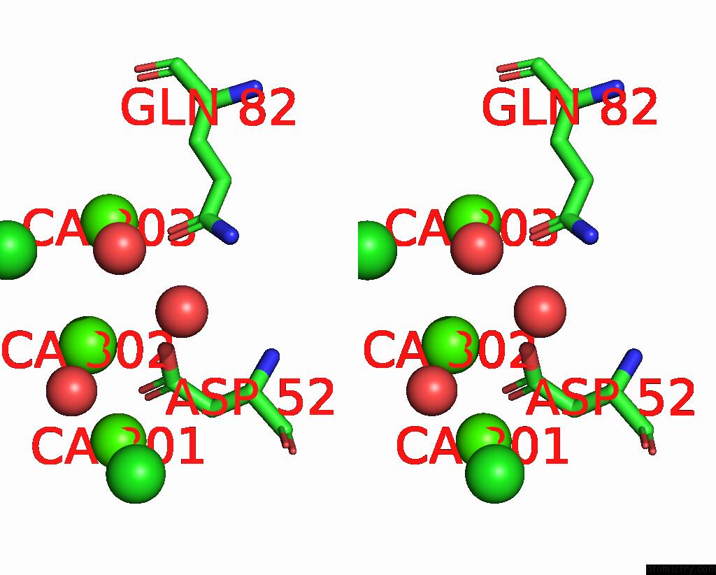
Stereo pair view
Mono view
Stereo pair view
A full contact list of Calcium with other atoms in the Ca binding
site number 2 of Crystal Structure of An 8-Repeat Consensus Tpr Superhelix in Tris Buffer with Calcium. within 5.0Å range:
|
Calcium binding site 3 out of 6 in 8cig
Go back to
Calcium binding site 3 out
of 6 in the Crystal Structure of An 8-Repeat Consensus Tpr Superhelix in Tris Buffer with Calcium.
Mono view
Stereo pair view
Mono view
Stereo pair view
A full contact list of Calcium with other atoms in the Ca binding
site number 3 of Crystal Structure of An 8-Repeat Consensus Tpr Superhelix in Tris Buffer with Calcium. within 5.0Å range:
|
Calcium binding site 4 out of 6 in 8cig
Go back to
Calcium binding site 4 out
of 6 in the Crystal Structure of An 8-Repeat Consensus Tpr Superhelix in Tris Buffer with Calcium.
Mono view
Stereo pair view
Mono view
Stereo pair view
A full contact list of Calcium with other atoms in the Ca binding
site number 4 of Crystal Structure of An 8-Repeat Consensus Tpr Superhelix in Tris Buffer with Calcium. within 5.0Å range:
|
Calcium binding site 5 out of 6 in 8cig
Go back to
Calcium binding site 5 out
of 6 in the Crystal Structure of An 8-Repeat Consensus Tpr Superhelix in Tris Buffer with Calcium.
Mono view
Stereo pair view
Mono view
Stereo pair view
A full contact list of Calcium with other atoms in the Ca binding
site number 5 of Crystal Structure of An 8-Repeat Consensus Tpr Superhelix in Tris Buffer with Calcium. within 5.0Å range:
|
Calcium binding site 6 out of 6 in 8cig
Go back to
Calcium binding site 6 out
of 6 in the Crystal Structure of An 8-Repeat Consensus Tpr Superhelix in Tris Buffer with Calcium.
Mono view
Stereo pair view
Mono view
Stereo pair view
A full contact list of Calcium with other atoms in the Ca binding
site number 6 of Crystal Structure of An 8-Repeat Consensus Tpr Superhelix in Tris Buffer with Calcium. within 5.0Å range:
|
Reference:
M.Liutkus,
A.L.Rojas,
I.R.Sasselli,
A.L.Cortajarena.
Diverse Crystalline Protein Scaffolds Through Metal-Dependent Polymorphism. To Be Published.
Page generated: Thu Jul 10 03:40:48 2025
Last articles
F in 7M03F in 7M02
F in 7LZU
F in 7LZW
F in 7LY8
F in 7LWG
F in 7LZV
F in 7LZF
F in 7LZD
F in 7LZA