Calcium »
PDB 8e51-8emw »
8e51 »
Calcium in PDB 8e51: Crystal Structure of Iridescent Shark Catfish Cadherin-23 EC1-2 and Protocadherin-15 EC1-2
Protein crystallography data
The structure of Crystal Structure of Iridescent Shark Catfish Cadherin-23 EC1-2 and Protocadherin-15 EC1-2, PDB code: 8e51
was solved by
E.Scheib,
C.R.Nisler,
M.Sotomayor,
with X-Ray Crystallography technique. A brief refinement statistics is given in the table below:
| Resolution Low / High (Å) | 44.60 / 2.59 |
| Space group | P 32 2 1 |
| Cell size a, b, c (Å), α, β, γ (°) | 75.816, 75.816, 242.68, 90, 90, 120 |
| R / Rfree (%) | 20.2 / 24.2 |
Other elements in 8e51:
The structure of Crystal Structure of Iridescent Shark Catfish Cadherin-23 EC1-2 and Protocadherin-15 EC1-2 also contains other interesting chemical elements:
| Potassium | (K) | 2 atoms |
Calcium Binding Sites:
Pages:
>>> Page 1 <<< Page 2, Binding sites: 11 - 13;Binding sites:
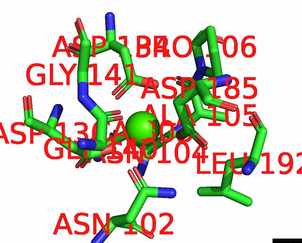
The binding sites of Calcium atom in the Crystal Structure of Iridescent Shark Catfish Cadherin-23 EC1-2 and Protocadherin-15 EC1-2 (pdb code 8e51). This binding sites where shown within 5.0 Angstroms radius around Calcium atom.In total 13 binding sites of Calcium where determined in the Crystal Structure of Iridescent Shark Catfish Cadherin-23 EC1-2 and Protocadherin-15 EC1-2, PDB code: 8e51:
Jump to Calcium binding site number: 1; 2; 3; 4; 5; 6; 7; 8; 9; 10;
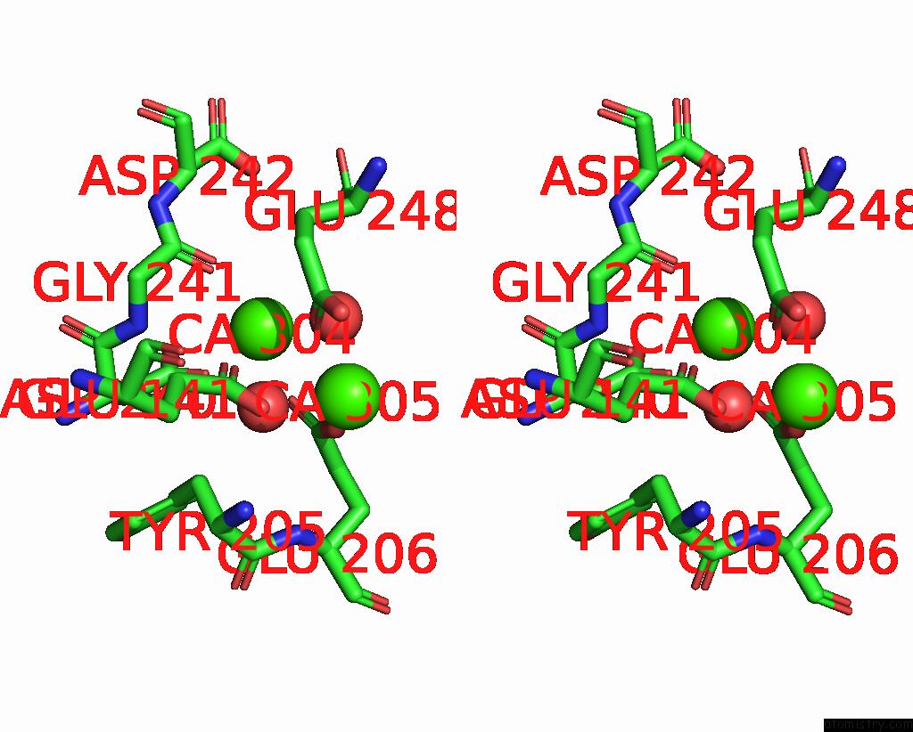
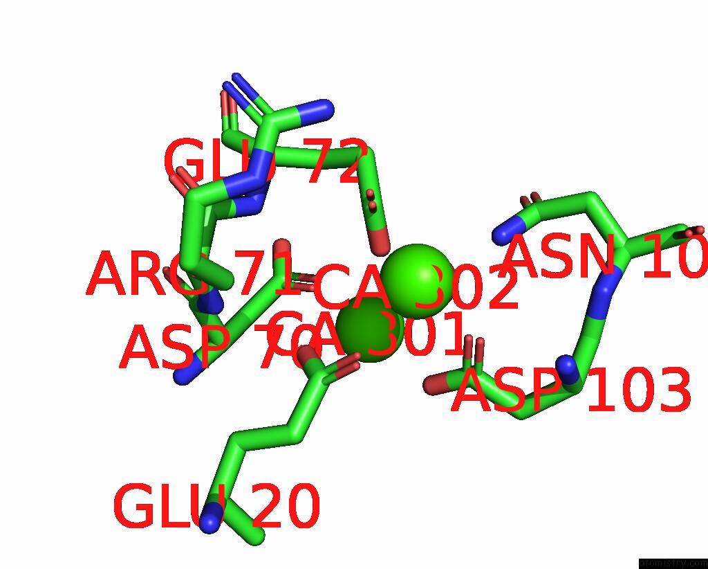
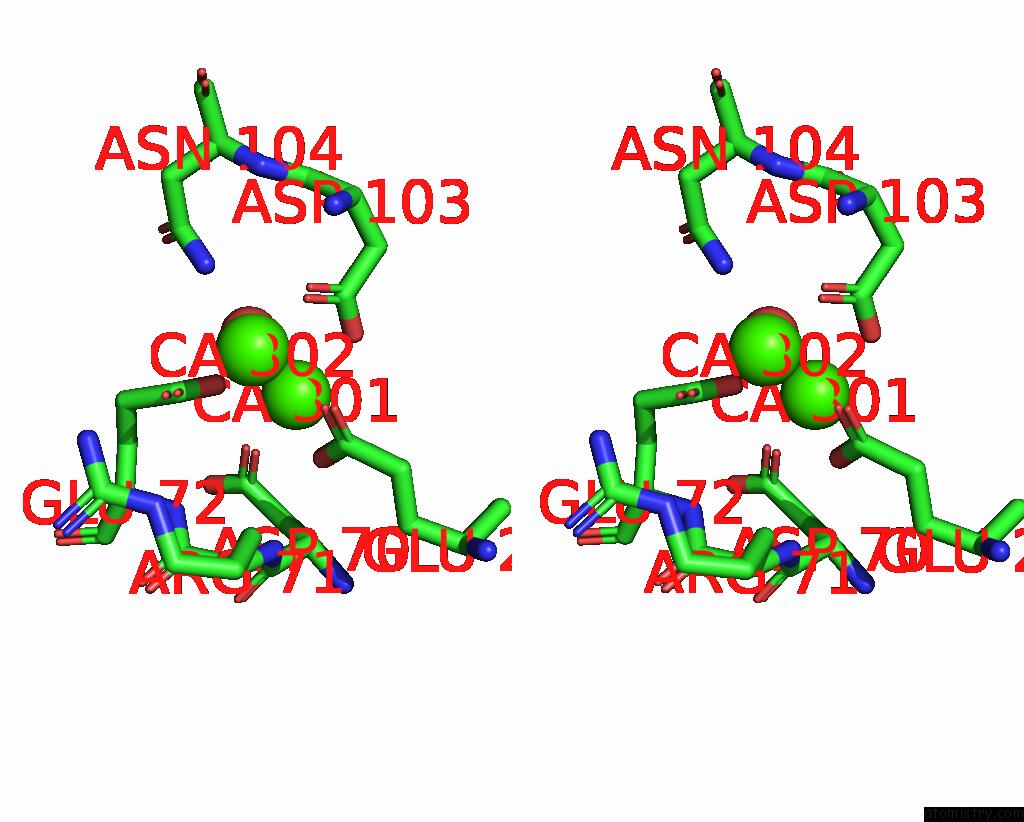
Calcium binding site 1 out of 13 in 8e51
Go back to
Calcium binding site 1 out
of 13 in the Crystal Structure of Iridescent Shark Catfish Cadherin-23 EC1-2 and Protocadherin-15 EC1-2
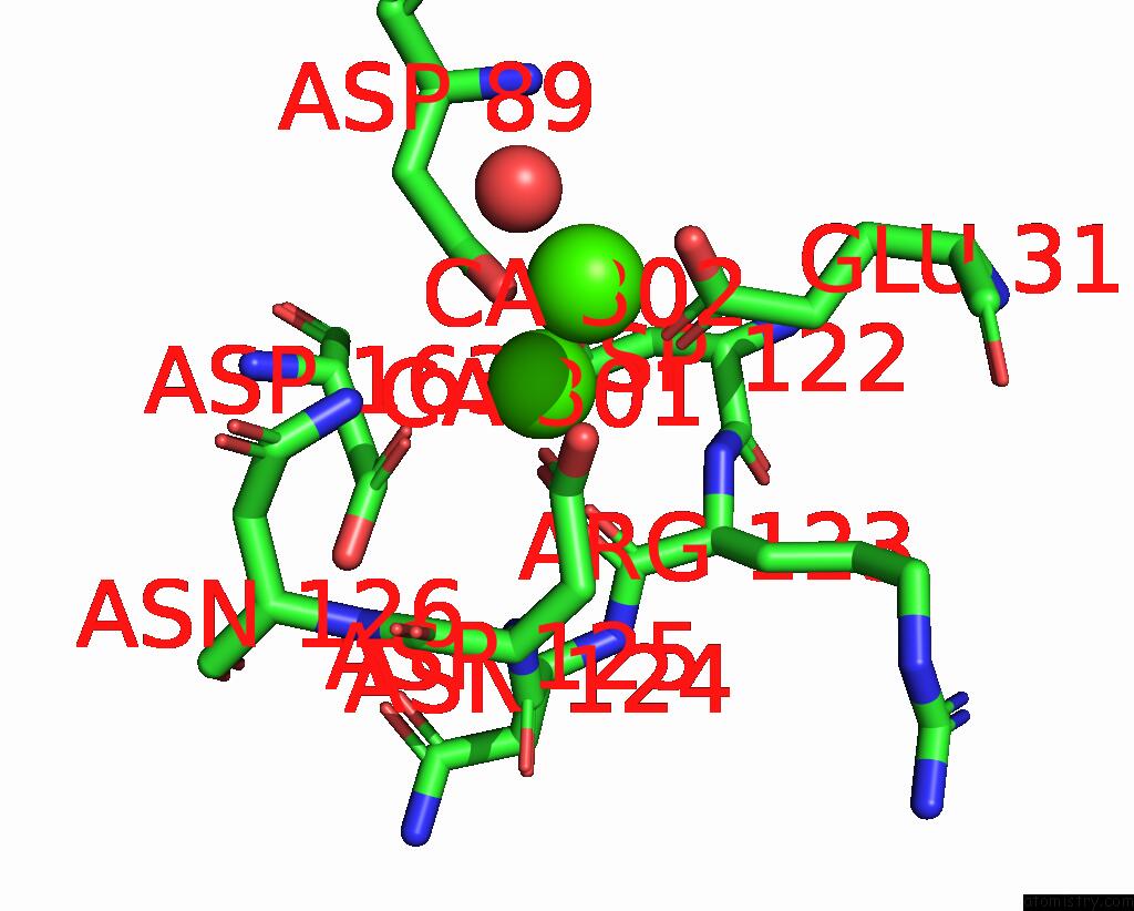
Mono view

Stereo pair view
Mono view
Stereo pair view
A full contact list of Calcium with other atoms in the Ca binding
site number 1 of Crystal Structure of Iridescent Shark Catfish Cadherin-23 EC1-2 and Protocadherin-15 EC1-2 within 5.0Å range:
|
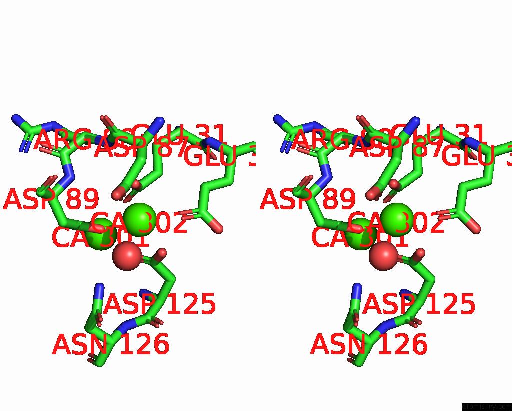
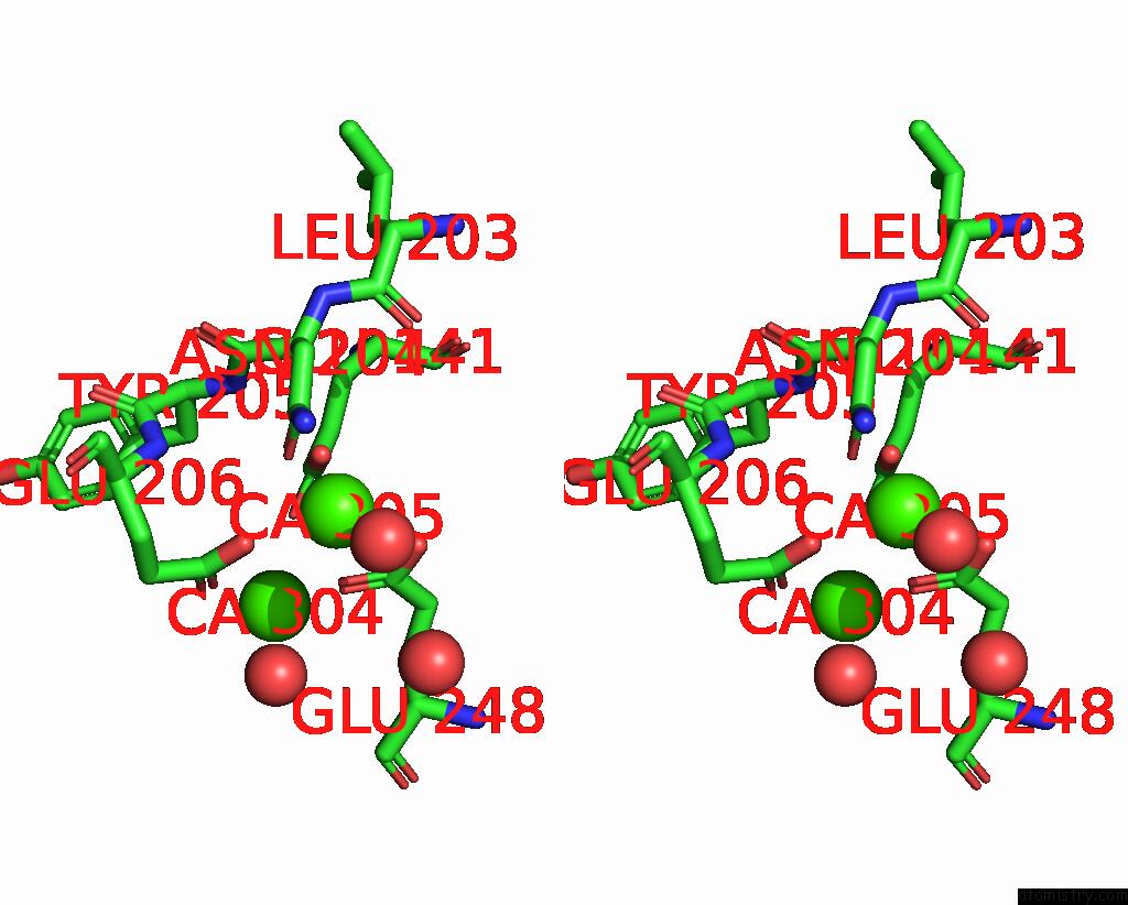
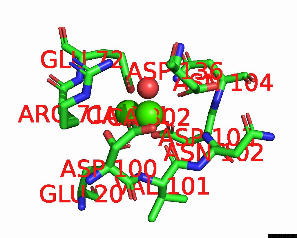
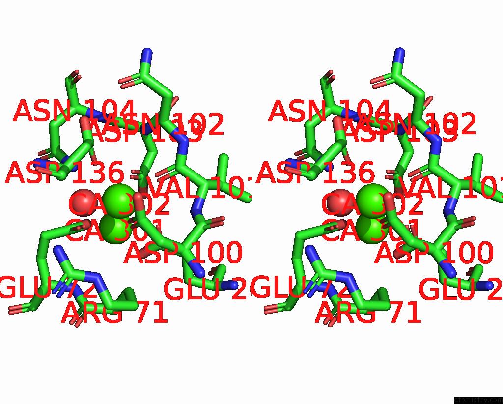
Calcium binding site 2 out of 13 in 8e51
Go back to
Calcium binding site 2 out
of 13 in the Crystal Structure of Iridescent Shark Catfish Cadherin-23 EC1-2 and Protocadherin-15 EC1-2
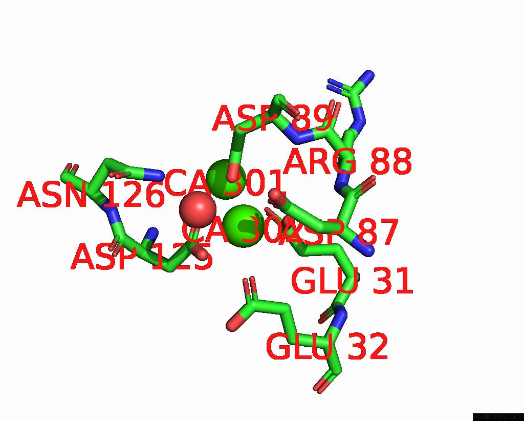
Mono view
Stereo pair view
Mono view
Stereo pair view
A full contact list of Calcium with other atoms in the Ca binding
site number 2 of Crystal Structure of Iridescent Shark Catfish Cadherin-23 EC1-2 and Protocadherin-15 EC1-2 within 5.0Å range:
|
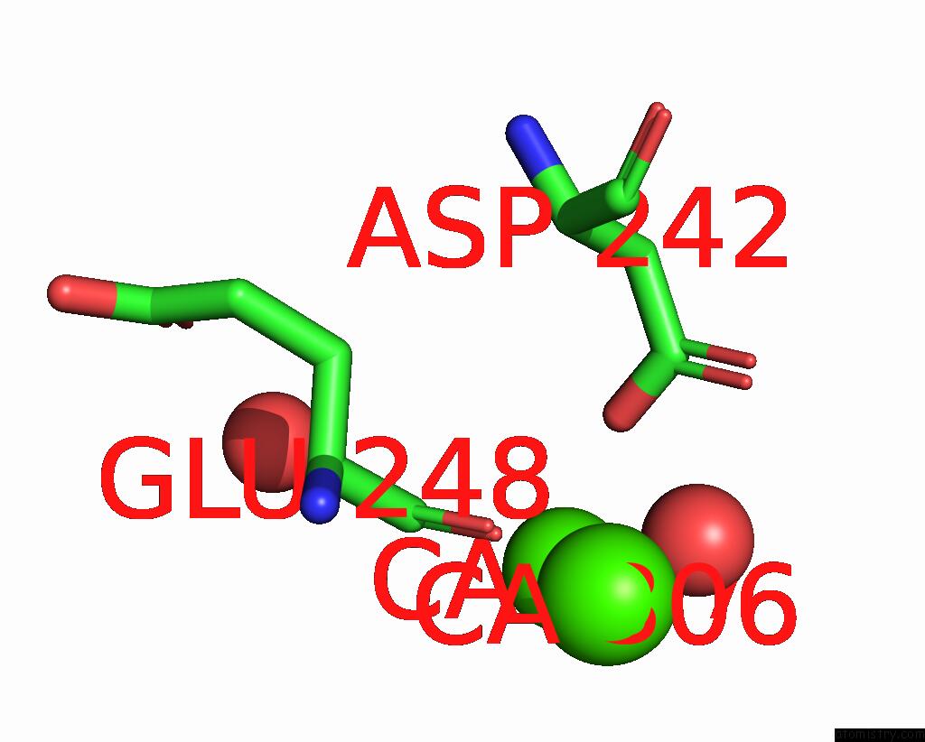
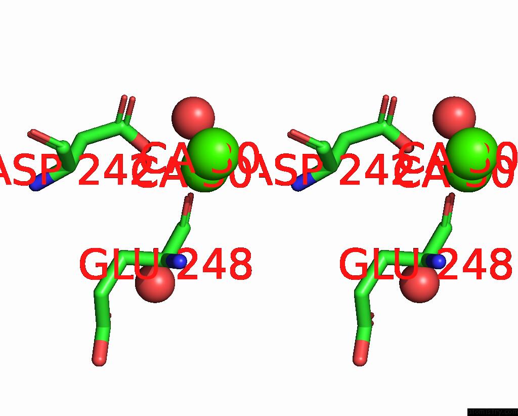
Calcium binding site 3 out of 13 in 8e51
Go back to
Calcium binding site 3 out
of 13 in the Crystal Structure of Iridescent Shark Catfish Cadherin-23 EC1-2 and Protocadherin-15 EC1-2
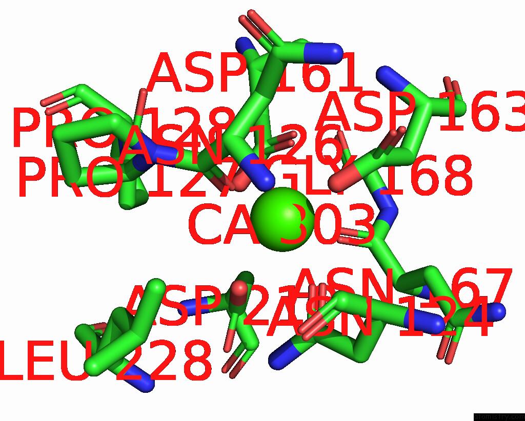
Mono view

Stereo pair view
Mono view
Stereo pair view
A full contact list of Calcium with other atoms in the Ca binding
site number 3 of Crystal Structure of Iridescent Shark Catfish Cadherin-23 EC1-2 and Protocadherin-15 EC1-2 within 5.0Å range:
|
Calcium binding site 4 out of 13 in 8e51
Go back to
Calcium binding site 4 out
of 13 in the Crystal Structure of Iridescent Shark Catfish Cadherin-23 EC1-2 and Protocadherin-15 EC1-2

Mono view
Stereo pair view
Mono view
Stereo pair view
A full contact list of Calcium with other atoms in the Ca binding
site number 4 of Crystal Structure of Iridescent Shark Catfish Cadherin-23 EC1-2 and Protocadherin-15 EC1-2 within 5.0Å range:
|
Calcium binding site 5 out of 13 in 8e51
Go back to
Calcium binding site 5 out
of 13 in the Crystal Structure of Iridescent Shark Catfish Cadherin-23 EC1-2 and Protocadherin-15 EC1-2

Mono view
Stereo pair view
Mono view
Stereo pair view
A full contact list of Calcium with other atoms in the Ca binding
site number 5 of Crystal Structure of Iridescent Shark Catfish Cadherin-23 EC1-2 and Protocadherin-15 EC1-2 within 5.0Å range:
|
Calcium binding site 6 out of 13 in 8e51
Go back to
Calcium binding site 6 out
of 13 in the Crystal Structure of Iridescent Shark Catfish Cadherin-23 EC1-2 and Protocadherin-15 EC1-2

Mono view

Stereo pair view
Mono view
Stereo pair view
A full contact list of Calcium with other atoms in the Ca binding
site number 6 of Crystal Structure of Iridescent Shark Catfish Cadherin-23 EC1-2 and Protocadherin-15 EC1-2 within 5.0Å range:
|
Calcium binding site 7 out of 13 in 8e51
Go back to
Calcium binding site 7 out
of 13 in the Crystal Structure of Iridescent Shark Catfish Cadherin-23 EC1-2 and Protocadherin-15 EC1-2
Mono view
Stereo pair view
Mono view
Stereo pair view
A full contact list of Calcium with other atoms in the Ca binding
site number 7 of Crystal Structure of Iridescent Shark Catfish Cadherin-23 EC1-2 and Protocadherin-15 EC1-2 within 5.0Å range:
|
Calcium binding site 8 out of 13 in 8e51
Go back to
Calcium binding site 8 out
of 13 in the Crystal Structure of Iridescent Shark Catfish Cadherin-23 EC1-2 and Protocadherin-15 EC1-2
Mono view
Stereo pair view
Mono view
Stereo pair view
A full contact list of Calcium with other atoms in the Ca binding
site number 8 of Crystal Structure of Iridescent Shark Catfish Cadherin-23 EC1-2 and Protocadherin-15 EC1-2 within 5.0Å range:
|
Calcium binding site 9 out of 13 in 8e51
Go back to
Calcium binding site 9 out
of 13 in the Crystal Structure of Iridescent Shark Catfish Cadherin-23 EC1-2 and Protocadherin-15 EC1-2
Mono view
Stereo pair view
Mono view
Stereo pair view
A full contact list of Calcium with other atoms in the Ca binding
site number 9 of Crystal Structure of Iridescent Shark Catfish Cadherin-23 EC1-2 and Protocadherin-15 EC1-2 within 5.0Å range:
|
Calcium binding site 10 out of 13 in 8e51
Go back to
Calcium binding site 10 out
of 13 in the Crystal Structure of Iridescent Shark Catfish Cadherin-23 EC1-2 and Protocadherin-15 EC1-2
Mono view

Stereo pair view
Mono view
Stereo pair view
A full contact list of Calcium with other atoms in the Ca binding
site number 10 of Crystal Structure of Iridescent Shark Catfish Cadherin-23 EC1-2 and Protocadherin-15 EC1-2 within 5.0Å range:
|
Reference:
C.R.Nisler,
Y.Narui,
E.Scheib,
D.Choudhary,
J.D.Bowman,
H.Mandayam Bharathi,
V.J.Lynch,
M.Sotomayor.
Interpreting the Evolutionary Echoes of A Protein Complex Essential For Inner-Ear Mechanosensation. Mol.Biol.Evol. 2023.
ISSN: ESSN 1537-1719
PubMed: 36911992
DOI: 10.1093/MOLBEV/MSAD057
Page generated: Thu Jul 10 04:02:56 2025
ISSN: ESSN 1537-1719
PubMed: 36911992
DOI: 10.1093/MOLBEV/MSAD057
Last articles
Cl in 5GSUCl in 5GS8
Cl in 5GQQ
Cl in 5G6U
Cl in 5GQN
Cl in 5GQM
Cl in 5GQL
Cl in 5GQK
Cl in 5GQJ
Cl in 5GNK