Calcium »
PDB 8gq9-8hb5 »
8gqb »
Calcium in PDB 8gqb: Crystal Structure of Lasso Peptide Epimerase Mslh D11A Mutant
Protein crystallography data
The structure of Crystal Structure of Lasso Peptide Epimerase Mslh D11A Mutant, PDB code: 8gqb
was solved by
Y.Nakashima,
H.Morita,
with X-Ray Crystallography technique. A brief refinement statistics is given in the table below:
| Resolution Low / High (Å) | 44.94 / 2.41 |
| Space group | I 4 2 2 |
| Cell size a, b, c (Å), α, β, γ (°) | 127.115, 127.115, 170.648, 90, 90, 90 |
| R / Rfree (%) | 19.6 / 22.4 |
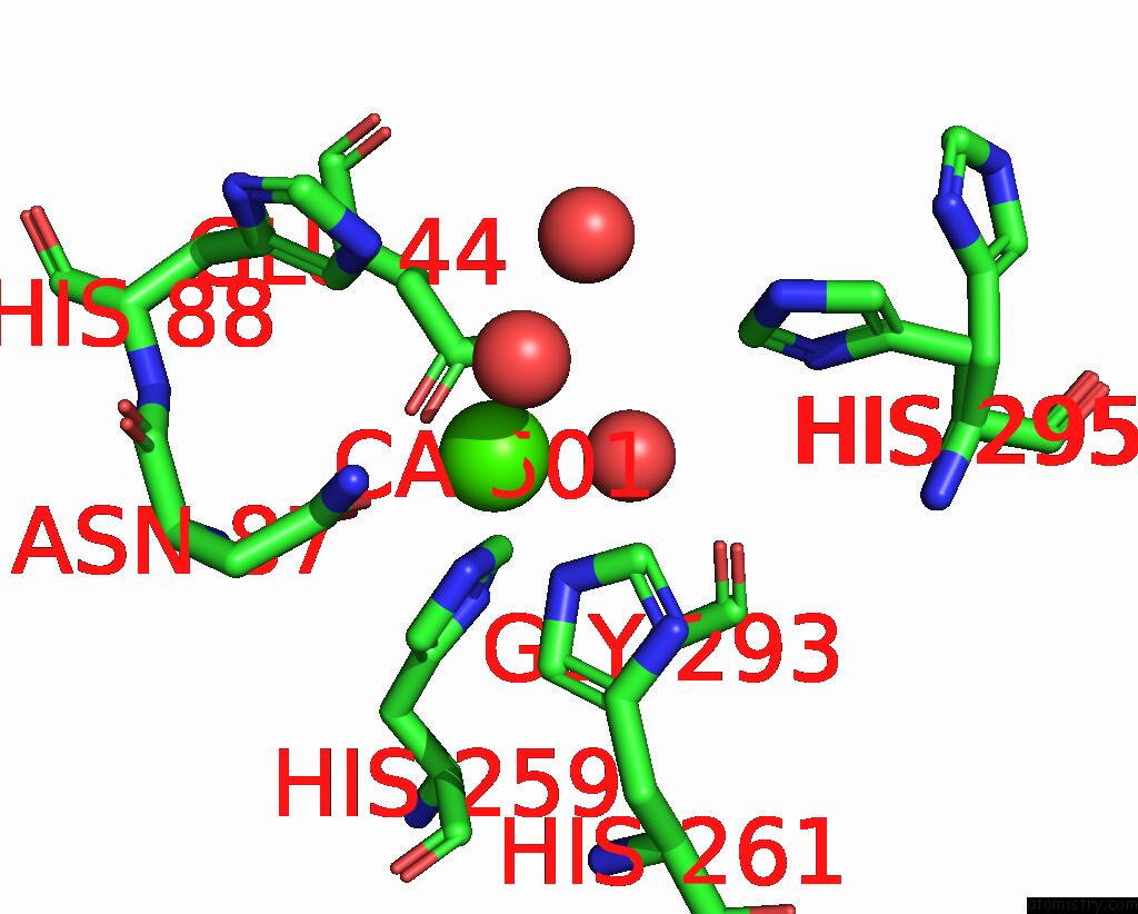
Calcium Binding Sites:
The binding sites of Calcium atom in the Crystal Structure of Lasso Peptide Epimerase Mslh D11A Mutant
(pdb code 8gqb). This binding sites where shown within
5.0 Angstroms radius around Calcium atom.
In total only one binding site of Calcium was determined in the Crystal Structure of Lasso Peptide Epimerase Mslh D11A Mutant, PDB code: 8gqb:
In total only one binding site of Calcium was determined in the Crystal Structure of Lasso Peptide Epimerase Mslh D11A Mutant, PDB code: 8gqb:
Calcium binding site 1 out of 1 in 8gqb
Go back to
Calcium binding site 1 out
of 1 in the Crystal Structure of Lasso Peptide Epimerase Mslh D11A Mutant
Mono view

Stereo pair view
Mono view
Stereo pair view
A full contact list of Calcium with other atoms in the Ca binding
site number 1 of Crystal Structure of Lasso Peptide Epimerase Mslh D11A Mutant within 5.0Å range:
|
Reference:
Y.Nakashima,
H.Morita.
X-Ray Crystallographic Analysis of Lasso Peptide Epimerase; Mslh To Be Published.
Page generated: Thu Jul 10 04:56:44 2025
Last articles
F in 4E1VF in 4DLE
F in 4E28
F in 4DZ9
F in 4E1N
F in 4DWV
F in 4DZ7
F in 4DXH
F in 4DXD
F in 4DVX