Calcium in PDB 8gt3: Crystal Structure of Human Cardiac Alpha Actin P109A Mutant (Adp-Pi State) in Complex with Fragmin F1 Domain
Protein crystallography data
The structure of Crystal Structure of Human Cardiac Alpha Actin P109A Mutant (Adp-Pi State) in Complex with Fragmin F1 Domain, PDB code: 8gt3
was solved by
M.Iwasa,
T.Oda,
S.Takeda,
with X-Ray Crystallography technique. A brief refinement statistics is given in the table below:
| Resolution Low / High (Å) | 45.55 / 1.50 |
| Space group | P 21 21 21 |
| Cell size a, b, c (Å), α, β, γ (°) | 57.171, 91.108, 115.142, 90, 90, 90 |
| R / Rfree (%) | 16.5 / 19.6 |
Other elements in 8gt3:
The structure of Crystal Structure of Human Cardiac Alpha Actin P109A Mutant (Adp-Pi State) in Complex with Fragmin F1 Domain also contains other interesting chemical elements:
| Magnesium | (Mg) | 1 atom |
Calcium Binding Sites:
The binding sites of Calcium atom in the Crystal Structure of Human Cardiac Alpha Actin P109A Mutant (Adp-Pi State) in Complex with Fragmin F1 Domain
(pdb code 8gt3). This binding sites where shown within
5.0 Angstroms radius around Calcium atom.
In total 2 binding sites of Calcium where determined in the Crystal Structure of Human Cardiac Alpha Actin P109A Mutant (Adp-Pi State) in Complex with Fragmin F1 Domain, PDB code: 8gt3:
Jump to Calcium binding site number: 1; 2;
In total 2 binding sites of Calcium where determined in the Crystal Structure of Human Cardiac Alpha Actin P109A Mutant (Adp-Pi State) in Complex with Fragmin F1 Domain, PDB code: 8gt3:
Jump to Calcium binding site number: 1; 2;
Calcium binding site 1 out of 2 in 8gt3
Go back to
Calcium binding site 1 out
of 2 in the Crystal Structure of Human Cardiac Alpha Actin P109A Mutant (Adp-Pi State) in Complex with Fragmin F1 Domain
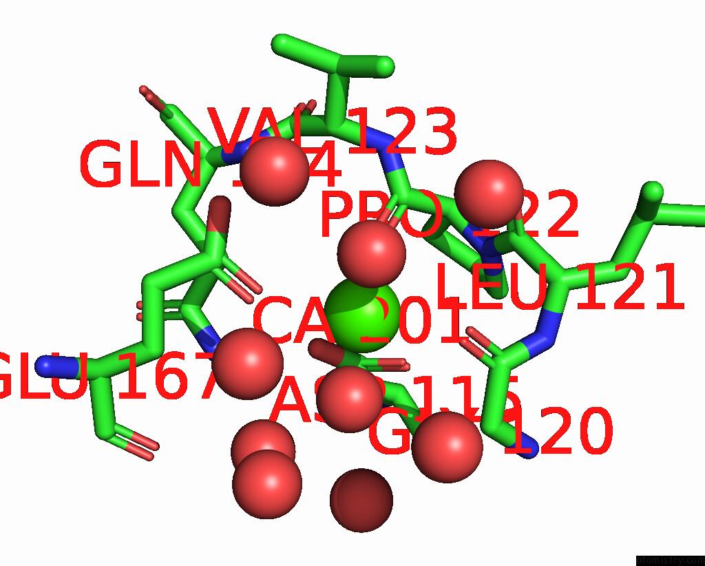
Mono view

Stereo pair view
Mono view
Stereo pair view
A full contact list of Calcium with other atoms in the Ca binding
site number 1 of Crystal Structure of Human Cardiac Alpha Actin P109A Mutant (Adp-Pi State) in Complex with Fragmin F1 Domain within 5.0Å range:
|
Calcium binding site 2 out of 2 in 8gt3
Go back to
Calcium binding site 2 out
of 2 in the Crystal Structure of Human Cardiac Alpha Actin P109A Mutant (Adp-Pi State) in Complex with Fragmin F1 Domain
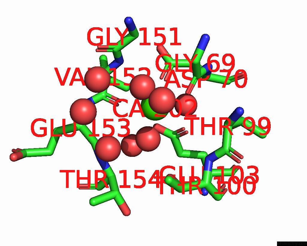
Mono view
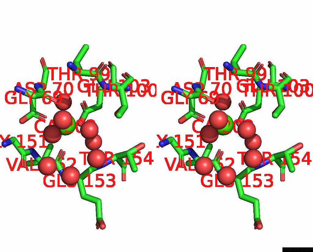
Stereo pair view
Mono view
Stereo pair view
A full contact list of Calcium with other atoms in the Ca binding
site number 2 of Crystal Structure of Human Cardiac Alpha Actin P109A Mutant (Adp-Pi State) in Complex with Fragmin F1 Domain within 5.0Å range:
|
Reference:
M.Iwasa,
S.Takeda,
A.Narita,
Y.Maeda,
T.Oda.
Mutagenic Analysis of Actin Reveals the Mechanism of HIS161 Flipping That Triggers Atp Hydrolysis Front Cell Dev Biol 2023.
ISSN: ESSN 2296-634X
DOI: 10.3389/FCELL.2023.1105460
Page generated: Fri Jul 19 09:17:35 2024
ISSN: ESSN 2296-634X
DOI: 10.3389/FCELL.2023.1105460
Last articles
Zn in 9MJ5Zn in 9HNW
Zn in 9G0L
Zn in 9FNE
Zn in 9DZN
Zn in 9E0I
Zn in 9D32
Zn in 9DAK
Zn in 8ZXC
Zn in 8ZUF