Calcium in PDB 8gyc: Annexin A5 Protein Dimer Mutant
Protein crystallography data
The structure of Annexin A5 Protein Dimer Mutant, PDB code: 8gyc
was solved by
Z.C.Hua,
W.Tang,
with X-Ray Crystallography technique. A brief refinement statistics is given in the table below:
Resolution Low / High (Å)
|
24.98 /
1.80
|
Space group
|
P 21 21 21
|
Cell size a, b, c (Å), α, β, γ (°)
|
69.198,
79.743,
133.936,
90,
90,
90
|
R / Rfree (%)
|
25.8 /
28.9
|
Calcium Binding Sites:
The binding sites of Calcium atom in the Annexin A5 Protein Dimer Mutant
(pdb code 8gyc). This binding sites where shown within
5.0 Angstroms radius around Calcium atom.
In total 7 binding sites of Calcium where determined in the
Annexin A5 Protein Dimer Mutant, PDB code: 8gyc:
Jump to Calcium binding site number:
1;
2;
3;
4;
5;
6;
7;
Calcium binding site 1 out
of 7 in 8gyc
Go back to
Calcium Binding Sites List in 8gyc
Calcium binding site 1 out
of 7 in the Annexin A5 Protein Dimer Mutant
Mono view
Stereo pair view
|
A full contact list of Calcium with other atoms in the Ca binding
site number 1 of Annexin A5 Protein Dimer Mutant within 5.0Å range:
probe
|
atom
|
residue
|
distance (Å)
|
B
|
Occ
|
A:Ca401
b:83.7
occ:1.00
|
OE1
|
A:GLU72
|
2.4
|
28.7
|
1.0
|
O
|
A:HOH562
|
2.5
|
37.2
|
1.0
|
O
|
A:HOH501
|
2.6
|
13.1
|
1.0
|
O
|
A:HOH724
|
2.7
|
40.3
|
1.0
|
O
|
A:HOH723
|
2.8
|
28.6
|
1.0
|
O
|
A:MET28
|
3.2
|
23.0
|
1.0
|
CD
|
A:GLU72
|
3.3
|
30.8
|
1.0
|
O
|
A:GLY30
|
3.4
|
23.4
|
1.0
|
O
|
A:HOH532
|
3.4
|
37.1
|
1.0
|
OE2
|
A:GLU72
|
3.5
|
28.3
|
1.0
|
C
|
A:MET28
|
4.4
|
21.6
|
1.0
|
C
|
A:GLY30
|
4.6
|
21.6
|
1.0
|
CG
|
A:GLU72
|
4.7
|
31.1
|
1.0
|
CB
|
A:MET28
|
4.7
|
26.5
|
1.0
|
N
|
A:GLY30
|
4.9
|
28.3
|
1.0
|
O
|
A:GLY32
|
5.0
|
22.1
|
1.0
|
|
Calcium binding site 2 out
of 7 in 8gyc
Go back to
Calcium Binding Sites List in 8gyc
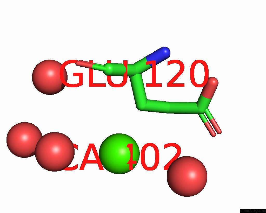
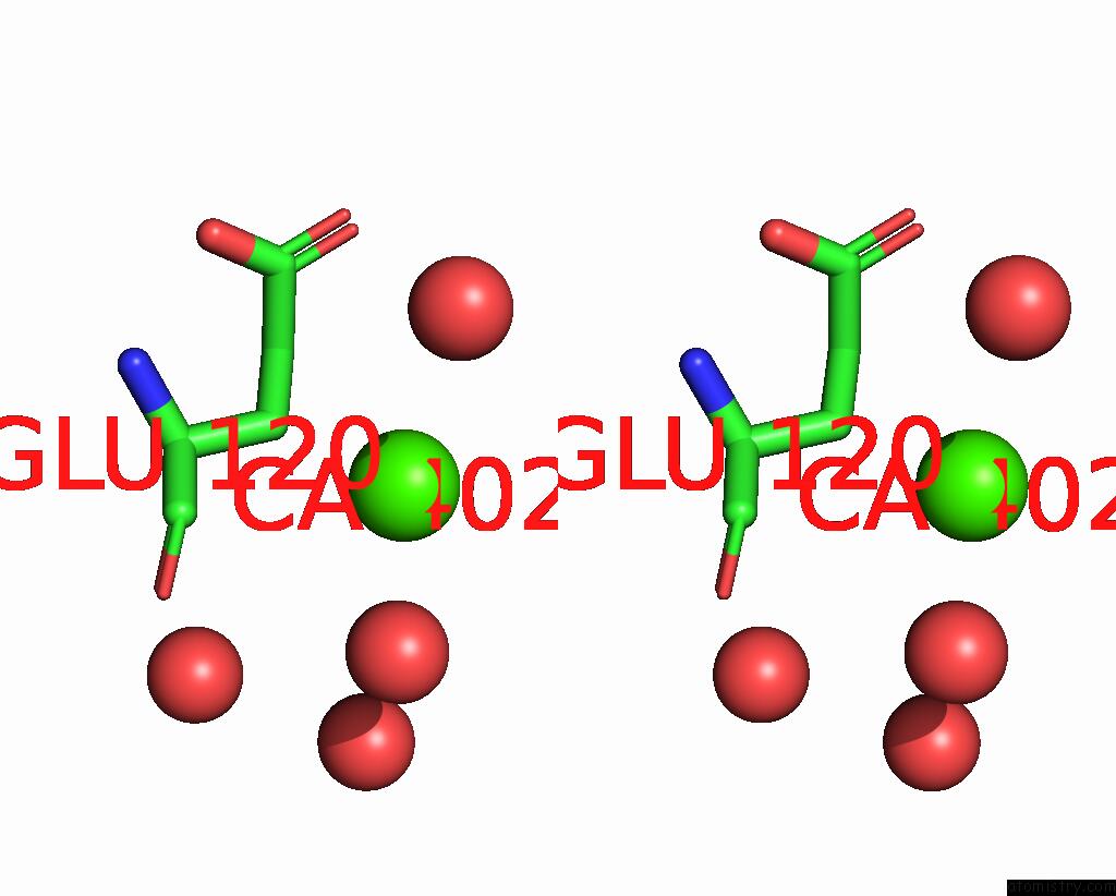
Calcium binding site 2 out
of 7 in the Annexin A5 Protein Dimer Mutant
Mono view
Stereo pair view
|
A full contact list of Calcium with other atoms in the Ca binding
site number 2 of Annexin A5 Protein Dimer Mutant within 5.0Å range:
probe
|
atom
|
residue
|
distance (Å)
|
B
|
Occ
|
A:Ca402
b:84.5
occ:1.00
|
CB
|
A:GLU120
|
2.8
|
38.0
|
1.0
|
CG
|
A:GLU120
|
3.0
|
52.9
|
1.0
|
CA
|
A:GLU120
|
4.1
|
29.4
|
1.0
|
O
|
A:HOH774
|
4.2
|
44.1
|
1.0
|
CD
|
A:GLU120
|
4.2
|
55.4
|
1.0
|
O
|
A:HOH734
|
4.2
|
40.5
|
1.0
|
OE1
|
A:GLU120
|
4.4
|
66.3
|
1.0
|
O
|
A:HOH575
|
4.6
|
33.3
|
1.0
|
C
|
A:GLU120
|
4.7
|
31.0
|
1.0
|
O
|
A:HOH535
|
4.9
|
41.1
|
1.0
|
|
Calcium binding site 3 out
of 7 in 8gyc
Go back to
Calcium Binding Sites List in 8gyc
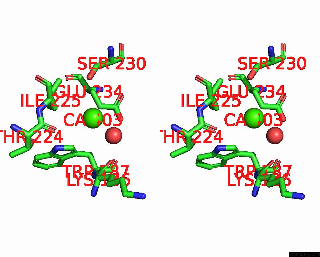
Calcium binding site 3 out
of 7 in the Annexin A5 Protein Dimer Mutant
Mono view
Stereo pair view
|
A full contact list of Calcium with other atoms in the Ca binding
site number 3 of Annexin A5 Protein Dimer Mutant within 5.0Å range:
probe
|
atom
|
residue
|
distance (Å)
|
B
|
Occ
|
A:Ca403
b:84.9
occ:1.00
|
O
|
A:HOH514
|
2.5
|
37.3
|
1.0
|
CG
|
A:GLU234
|
2.8
|
34.3
|
1.0
|
CB
|
A:GLU234
|
3.7
|
28.9
|
1.0
|
OG
|
A:SER230
|
3.7
|
56.0
|
1.0
|
O
|
A:THR224
|
3.8
|
40.1
|
1.0
|
CG1
|
A:ILE225
|
3.9
|
34.5
|
1.0
|
CD
|
A:GLU234
|
3.9
|
41.2
|
1.0
|
CD1
|
A:TRP187
|
4.0
|
30.1
|
1.0
|
OE2
|
A:GLU234
|
4.2
|
46.6
|
1.0
|
CA
|
A:ILE225
|
4.3
|
39.1
|
1.0
|
CB
|
A:ILE225
|
4.5
|
32.9
|
1.0
|
O
|
A:LYS186
|
4.5
|
30.7
|
1.0
|
CG2
|
A:ILE225
|
4.6
|
35.4
|
1.0
|
CB
|
A:SER230
|
4.7
|
51.0
|
1.0
|
OE1
|
A:GLU234
|
4.8
|
37.0
|
1.0
|
NE1
|
A:TRP187
|
4.8
|
28.6
|
1.0
|
C
|
A:THR224
|
4.8
|
40.5
|
1.0
|
CD1
|
A:ILE225
|
5.0
|
26.8
|
1.0
|
CG
|
A:TRP187
|
5.0
|
26.1
|
1.0
|
|
Calcium binding site 4 out
of 7 in 8gyc
Go back to
Calcium Binding Sites List in 8gyc
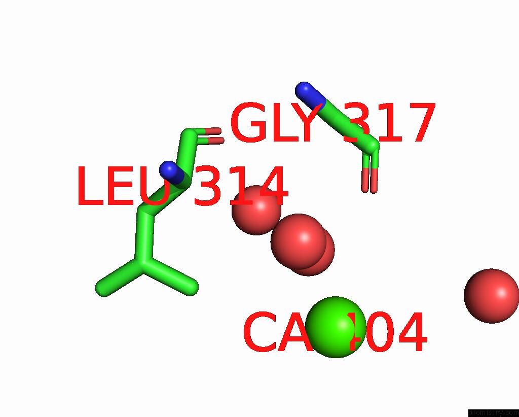
Calcium binding site 4 out
of 7 in the Annexin A5 Protein Dimer Mutant
Mono view
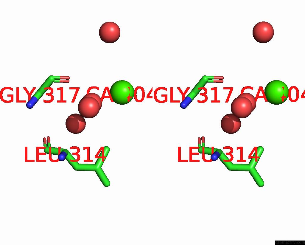
Stereo pair view
|
A full contact list of Calcium with other atoms in the Ca binding
site number 4 of Annexin A5 Protein Dimer Mutant within 5.0Å range:
probe
|
atom
|
residue
|
distance (Å)
|
B
|
Occ
|
A:Ca404
b:79.6
occ:1.00
|
O
|
A:HOH511
|
2.5
|
38.4
|
1.0
|
O
|
A:HOH692
|
4.0
|
33.2
|
1.0
|
O
|
A:GLY317
|
4.3
|
21.8
|
1.0
|
O
|
A:HOH604
|
4.4
|
26.7
|
1.0
|
CD2
|
A:LEU314
|
4.6
|
18.8
|
1.0
|
O
|
A:HOH771
|
4.7
|
43.8
|
1.0
|
|
Calcium binding site 5 out
of 7 in 8gyc
Go back to
Calcium Binding Sites List in 8gyc
Calcium binding site 5 out
of 7 in the Annexin A5 Protein Dimer Mutant
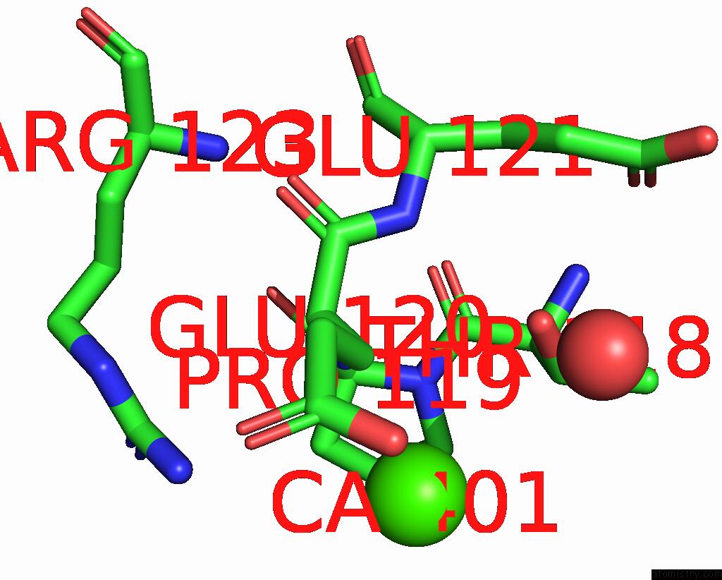
Mono view
Stereo pair view
|
A full contact list of Calcium with other atoms in the Ca binding
site number 5 of Annexin A5 Protein Dimer Mutant within 5.0Å range:
probe
|
atom
|
residue
|
distance (Å)
|
B
|
Occ
|
B:Ca401
b:88.2
occ:1.00
|
CB
|
B:GLU120
|
2.8
|
39.1
|
1.0
|
N
|
B:GLU120
|
3.0
|
25.9
|
1.0
|
CA
|
B:GLU120
|
3.5
|
30.7
|
1.0
|
OG1
|
B:THR118
|
3.9
|
22.3
|
1.0
|
OE2
|
B:GLU120
|
4.0
|
65.5
|
1.0
|
CG
|
B:GLU120
|
4.0
|
55.7
|
1.0
|
CB
|
B:THR118
|
4.1
|
28.6
|
1.0
|
CD
|
B:GLU120
|
4.1
|
68.3
|
1.0
|
C
|
B:PRO119
|
4.1
|
22.8
|
1.0
|
O
|
B:HOH510
|
4.1
|
38.1
|
1.0
|
N
|
B:PRO119
|
4.2
|
28.9
|
1.0
|
NH2
|
B:ARG123
|
4.3
|
40.0
|
1.0
|
CD
|
B:PRO119
|
4.3
|
29.6
|
1.0
|
CB
|
B:PRO119
|
4.3
|
31.0
|
1.0
|
CA
|
B:PRO119
|
4.5
|
23.0
|
1.0
|
C
|
B:THR118
|
4.7
|
25.4
|
1.0
|
C
|
B:GLU120
|
4.8
|
33.1
|
1.0
|
CG
|
B:PRO119
|
4.8
|
30.7
|
1.0
|
OE1
|
B:GLU120
|
4.8
|
70.1
|
1.0
|
N
|
B:GLU121
|
5.0
|
24.7
|
1.0
|
|
Calcium binding site 6 out
of 7 in 8gyc
Go back to
Calcium Binding Sites List in 8gyc
Calcium binding site 6 out
of 7 in the Annexin A5 Protein Dimer Mutant
Mono view
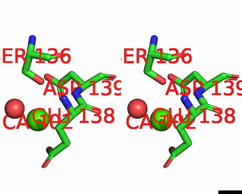
Stereo pair view
|
A full contact list of Calcium with other atoms in the Ca binding
site number 6 of Annexin A5 Protein Dimer Mutant within 5.0Å range:
probe
|
atom
|
residue
|
distance (Å)
|
B
|
Occ
|
B:Ca402
b:72.8
occ:1.00
|
O
|
B:HOH709
|
2.5
|
44.8
|
1.0
|
OD1
|
B:ASP139
|
2.6
|
39.8
|
1.0
|
OE2
|
B:GLU138
|
2.9
|
49.2
|
1.0
|
CG
|
B:ASP139
|
3.3
|
35.0
|
1.0
|
OD2
|
B:ASP139
|
3.6
|
30.1
|
1.0
|
CD
|
B:GLU138
|
3.9
|
36.5
|
1.0
|
OG
|
B:SER136
|
4.1
|
31.2
|
1.0
|
CG
|
B:GLU138
|
4.4
|
33.4
|
1.0
|
N
|
B:ASP139
|
4.5
|
24.2
|
1.0
|
CB
|
B:ASP139
|
4.6
|
28.2
|
1.0
|
CB
|
B:GLU138
|
4.6
|
29.3
|
1.0
|
CA
|
B:ASP139
|
4.8
|
24.9
|
1.0
|
OE1
|
B:GLU138
|
4.9
|
33.8
|
1.0
|
|
Calcium binding site 7 out
of 7 in 8gyc
Go back to
Calcium Binding Sites List in 8gyc
Calcium binding site 7 out
of 7 in the Annexin A5 Protein Dimer Mutant
Mono view
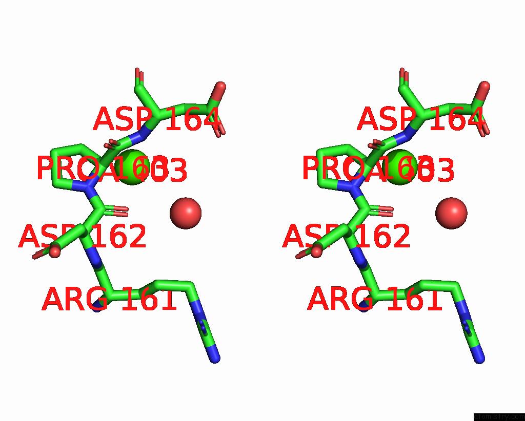
Stereo pair view
|
A full contact list of Calcium with other atoms in the Ca binding
site number 7 of Annexin A5 Protein Dimer Mutant within 5.0Å range:
probe
|
atom
|
residue
|
distance (Å)
|
B
|
Occ
|
B:Ca403
b:92.0
occ:1.00
|
CA
|
B:PRO163
|
2.8
|
55.1
|
1.0
|
CB
|
B:PRO163
|
3.2
|
62.6
|
1.0
|
N
|
B:ASP164
|
3.4
|
60.4
|
1.0
|
C
|
B:PRO163
|
3.7
|
52.4
|
1.0
|
N
|
B:PRO163
|
3.9
|
52.2
|
1.0
|
O
|
B:ASP162
|
4.1
|
40.0
|
1.0
|
O
|
B:HOH678
|
4.4
|
39.6
|
1.0
|
C
|
B:ASP162
|
4.4
|
41.9
|
1.0
|
CG
|
B:PRO163
|
4.6
|
57.4
|
1.0
|
CA
|
B:ASP164
|
4.7
|
58.2
|
1.0
|
CD
|
B:PRO163
|
4.8
|
53.5
|
1.0
|
O
|
B:PRO163
|
4.9
|
50.9
|
1.0
|
CB
|
B:ASP164
|
4.9
|
69.1
|
1.0
|
O
|
B:ARG161
|
5.0
|
45.3
|
1.0
|
|
Reference:
Z.C.Hua,
W.Tang.
Structure Dissection of the Membrane Aggregation Mechanism Induced By Annexin A5 Mutation To Be Published.
Page generated: Fri Jul 19 09:19:50 2024
|