Calcium »
PDB 8hcj-8ic1 »
8hfm »
Calcium in PDB 8hfm: Crystal Structure of Mycobacterium Smegmatis Mshc
Enzymatic activity of Crystal Structure of Mycobacterium Smegmatis Mshc
All present enzymatic activity of Crystal Structure of Mycobacterium Smegmatis Mshc:
6.3.1.13;
6.3.1.13;
Protein crystallography data
The structure of Crystal Structure of Mycobacterium Smegmatis Mshc, PDB code: 8hfm
was solved by
L.Pang,
S.D.Weeks,
S.V.Strelkov,
with X-Ray Crystallography technique. A brief refinement statistics is given in the table below:
| Resolution Low / High (Å) | 48.32 / 2.41 |
| Space group | P 41 2 2 |
| Cell size a, b, c (Å), α, β, γ (°) | 69.81, 69.81, 236.011, 90, 90, 90 |
| R / Rfree (%) | 20.6 / 23.8 |
Other elements in 8hfm:
The structure of Crystal Structure of Mycobacterium Smegmatis Mshc also contains other interesting chemical elements:
| Zinc | (Zn) | 1 atom |
Calcium Binding Sites:
The binding sites of Calcium atom in the Crystal Structure of Mycobacterium Smegmatis Mshc
(pdb code 8hfm). This binding sites where shown within
5.0 Angstroms radius around Calcium atom.
In total only one binding site of Calcium was determined in the Crystal Structure of Mycobacterium Smegmatis Mshc, PDB code: 8hfm:
In total only one binding site of Calcium was determined in the Crystal Structure of Mycobacterium Smegmatis Mshc, PDB code: 8hfm:
Calcium binding site 1 out of 1 in 8hfm
Go back to
Calcium binding site 1 out
of 1 in the Crystal Structure of Mycobacterium Smegmatis Mshc
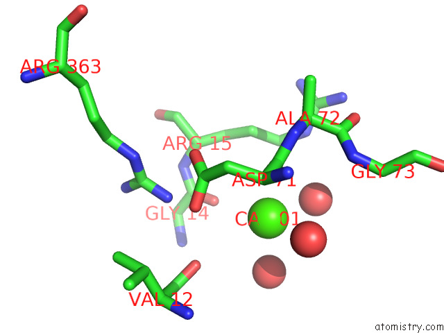
Mono view
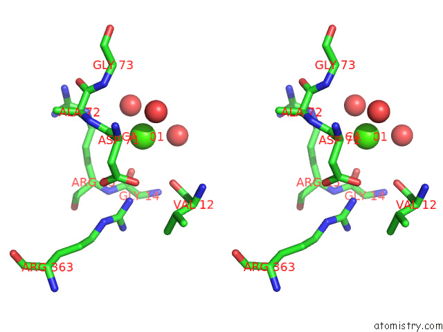
Stereo pair view
Mono view
Stereo pair view
A full contact list of Calcium with other atoms in the Ca binding
site number 1 of Crystal Structure of Mycobacterium Smegmatis Mshc within 5.0Å range:
|
Reference:
L.Pang,
S.Lenders,
E.M.Osipov,
S.D.Weeks,
J.Rozenski,
T.Piller,
D.Cappoen,
S.V.Strelkov,
A.Van Aerschot.
Structural Basis of Cysteine Ligase Mshc Inhibition By Cysteinyl-Sulfonamides. Int J Mol Sci V. 23 2022.
ISSN: ESSN 1422-0067
PubMed: 36499418
DOI: 10.3390/IJMS232315095
Page generated: Thu Jul 10 05:03:36 2025
ISSN: ESSN 1422-0067
PubMed: 36499418
DOI: 10.3390/IJMS232315095
Last articles
F in 7LGXF in 7LGK
F in 7LG8
F in 7LD3
F in 7LCR
F in 7LCM
F in 7LCO
F in 7LCK
F in 7LCJ
F in 7LCI