Calcium »
PDB 8hcj-8ic1 »
8i5p »
Calcium in PDB 8i5p: Crystal Structure of TXGH116 D593A Acid/Base Mutant From Thermoanaerobacterium Xylanolyticum with Cellobiose
Enzymatic activity of Crystal Structure of TXGH116 D593A Acid/Base Mutant From Thermoanaerobacterium Xylanolyticum with Cellobiose
All present enzymatic activity of Crystal Structure of TXGH116 D593A Acid/Base Mutant From Thermoanaerobacterium Xylanolyticum with Cellobiose:
3.2.1.21;
3.2.1.21;
Protein crystallography data
The structure of Crystal Structure of TXGH116 D593A Acid/Base Mutant From Thermoanaerobacterium Xylanolyticum with Cellobiose, PDB code: 8i5p
was solved by
S.Pengthaisong,
J.R.Ketudat Cairns,
with X-Ray Crystallography technique. A brief refinement statistics is given in the table below:
| Resolution Low / High (Å) | 40.00 / 2.35 |
| Space group | P 21 21 21 |
| Cell size a, b, c (Å), α, β, γ (°) | 98.072, 100.504, 173.783, 90, 90, 90 |
| R / Rfree (%) | 19 / 24.1 |
Calcium Binding Sites:
The binding sites of Calcium atom in the Crystal Structure of TXGH116 D593A Acid/Base Mutant From Thermoanaerobacterium Xylanolyticum with Cellobiose
(pdb code 8i5p). This binding sites where shown within
5.0 Angstroms radius around Calcium atom.
In total 2 binding sites of Calcium where determined in the Crystal Structure of TXGH116 D593A Acid/Base Mutant From Thermoanaerobacterium Xylanolyticum with Cellobiose, PDB code: 8i5p:
Jump to Calcium binding site number: 1; 2;
In total 2 binding sites of Calcium where determined in the Crystal Structure of TXGH116 D593A Acid/Base Mutant From Thermoanaerobacterium Xylanolyticum with Cellobiose, PDB code: 8i5p:
Jump to Calcium binding site number: 1; 2;
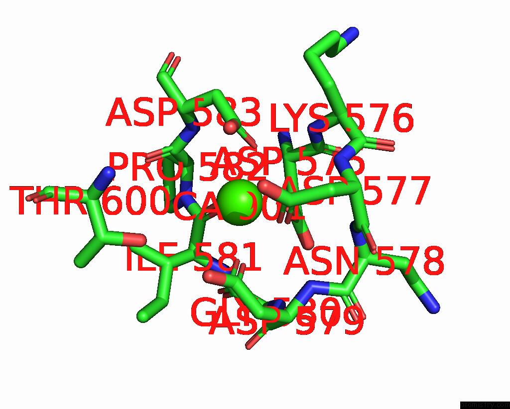
Calcium binding site 1 out of 2 in 8i5p
Go back to
Calcium binding site 1 out
of 2 in the Crystal Structure of TXGH116 D593A Acid/Base Mutant From Thermoanaerobacterium Xylanolyticum with Cellobiose

Mono view
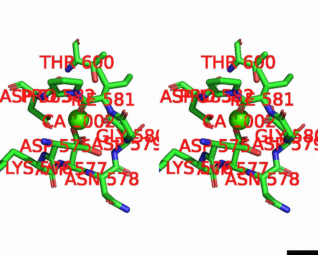
Stereo pair view
Mono view
Stereo pair view
A full contact list of Calcium with other atoms in the Ca binding
site number 1 of Crystal Structure of TXGH116 D593A Acid/Base Mutant From Thermoanaerobacterium Xylanolyticum with Cellobiose within 5.0Å range:
|
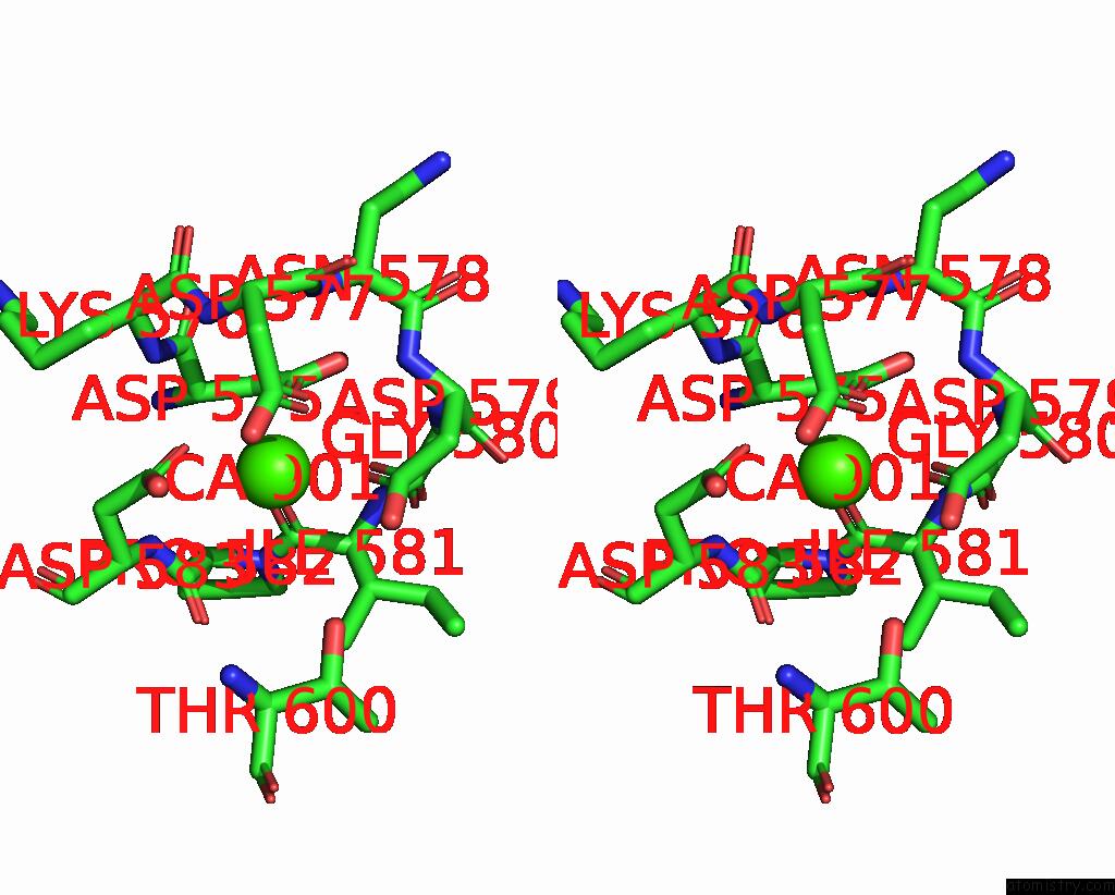
Calcium binding site 2 out of 2 in 8i5p
Go back to
Calcium binding site 2 out
of 2 in the Crystal Structure of TXGH116 D593A Acid/Base Mutant From Thermoanaerobacterium Xylanolyticum with Cellobiose
Mono view
Stereo pair view
Mono view
Stereo pair view
A full contact list of Calcium with other atoms in the Ca binding
site number 2 of Crystal Structure of TXGH116 D593A Acid/Base Mutant From Thermoanaerobacterium Xylanolyticum with Cellobiose within 5.0Å range:
|
Reference:
S.Pengthaisong,
B.Piniello,
G.J.Davies,
C.Rovira,
J.R.K.Cairns.
Reaction Mechanism of Glycoside Hydrolase Family 116 Utilizes Perpendicular Protonation Acs Catalysis 5850 2023.
ISSN: ESSN 2155-5435
DOI: 10.1021/ACSCATAL.3C00620
Page generated: Thu Jul 10 05:11:16 2025
ISSN: ESSN 2155-5435
DOI: 10.1021/ACSCATAL.3C00620
Last articles
Cl in 5KOECl in 5KP1
Cl in 5KOP
Cl in 5KP3
Cl in 5KOR
Cl in 5KOL
Cl in 5KN1
Cl in 5KMU
Cl in 5KMD
Cl in 5KMB