Calcium »
PDB 8ihn-8izi »
8iw5 »
Calcium in PDB 8iw5: Crystal Structure of Liprin-Beta H2H3 Dimer
Protein crystallography data
The structure of Crystal Structure of Liprin-Beta H2H3 Dimer, PDB code: 8iw5
was solved by
J.Zhang,
S.Chen,
Z.Wei,
with X-Ray Crystallography technique. A brief refinement statistics is given in the table below:
| Resolution Low / High (Å) | 27.33 / 1.70 |
| Space group | P 1 21 1 |
| Cell size a, b, c (Å), α, β, γ (°) | 29.793, 50.637, 30.78, 90, 113.44, 90 |
| R / Rfree (%) | 22.1 / 27.2 |
Calcium Binding Sites:
The binding sites of Calcium atom in the Crystal Structure of Liprin-Beta H2H3 Dimer
(pdb code 8iw5). This binding sites where shown within
5.0 Angstroms radius around Calcium atom.
In total only one binding site of Calcium was determined in the Crystal Structure of Liprin-Beta H2H3 Dimer, PDB code: 8iw5:
In total only one binding site of Calcium was determined in the Crystal Structure of Liprin-Beta H2H3 Dimer, PDB code: 8iw5:
Calcium binding site 1 out of 1 in 8iw5
Go back to
Calcium binding site 1 out
of 1 in the Crystal Structure of Liprin-Beta H2H3 Dimer
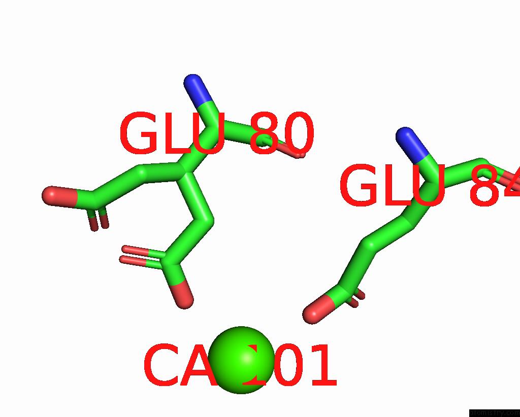
Mono view
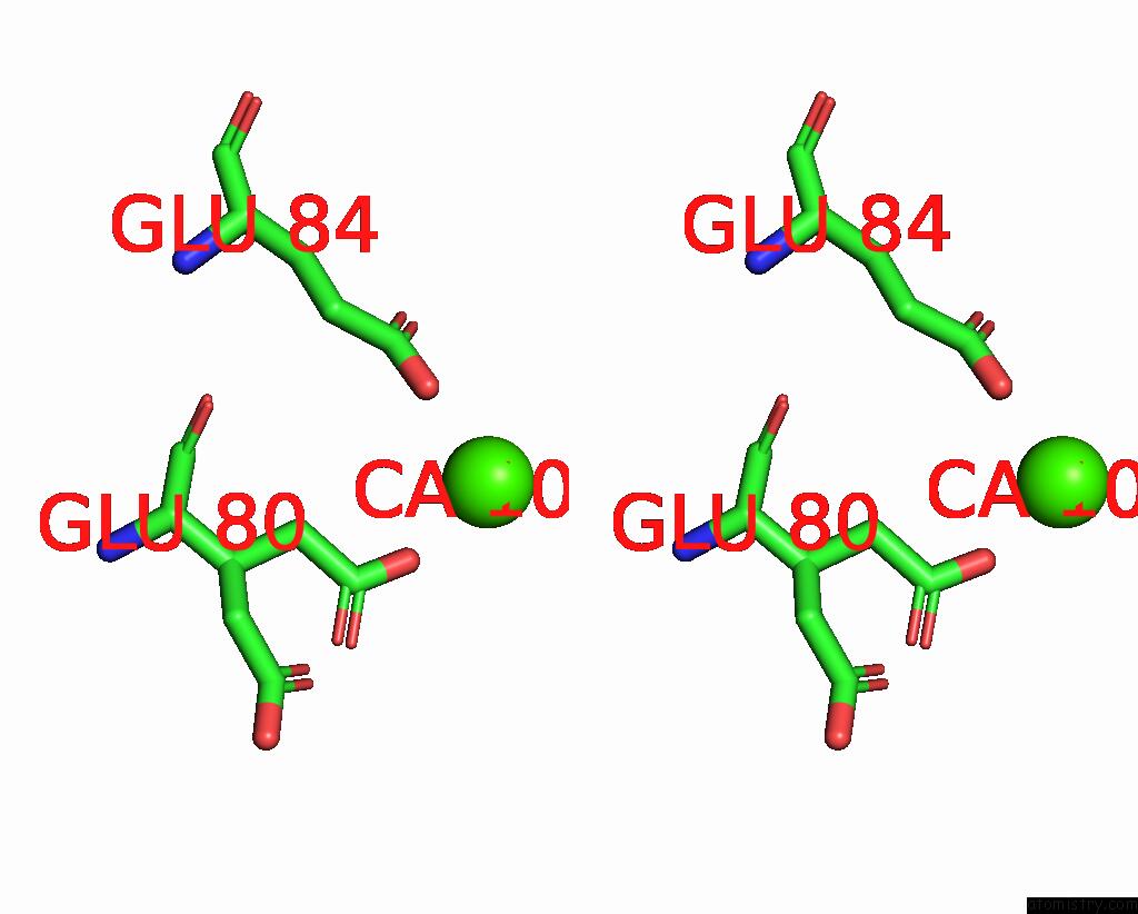
Stereo pair view
Mono view
Stereo pair view
A full contact list of Calcium with other atoms in the Ca binding
site number 1 of Crystal Structure of Liprin-Beta H2H3 Dimer within 5.0Å range:
|
Reference:
K.Guo,
J.Zhang,
P.Huang,
Y.Xu,
W.Pan,
K.Li,
L.Chen,
L.Luo,
W.Yu,
S.Chen,
S.He,
Z.Wei,
C.Yu.
KANK1 Shapes Focal Adhesions By Orchestrating Protein Binding, Mechanical Force Sensing, and Phase Separation. Cell Rep V. 42 13321 2023.
ISSN: ESSN 2211-1247
PubMed: 37874676
DOI: 10.1016/J.CELREP.2023.113321
Page generated: Fri Jul 19 09:53:35 2024
ISSN: ESSN 2211-1247
PubMed: 37874676
DOI: 10.1016/J.CELREP.2023.113321
Last articles
Zn in 9JYWZn in 9IR4
Zn in 9IR3
Zn in 9GMX
Zn in 9GMW
Zn in 9JEJ
Zn in 9ERF
Zn in 9ERE
Zn in 9EGV
Zn in 9EGW