Calcium »
PDB 8izk-8jx8 »
8jbo »
Calcium in PDB 8jbo: Crystal Structure of TXGH116 From Thermoanaerobacterium Xylanolyticum with Isofagomine
Protein crystallography data
The structure of Crystal Structure of TXGH116 From Thermoanaerobacterium Xylanolyticum with Isofagomine, PDB code: 8jbo
was solved by
S.Pengthaisong,
J.R.Ketudat Cairns,
with X-Ray Crystallography technique. A brief refinement statistics is given in the table below:
| Resolution Low / High (Å) | 50.01 / 2.00 |
| Space group | P 21 21 21 |
| Cell size a, b, c (Å), α, β, γ (°) | 54.791, 164.399, 179.614, 90, 90, 90 |
| R / Rfree (%) | 15.5 / 19.3 |
Calcium Binding Sites:
The binding sites of Calcium atom in the Crystal Structure of TXGH116 From Thermoanaerobacterium Xylanolyticum with Isofagomine
(pdb code 8jbo). This binding sites where shown within
5.0 Angstroms radius around Calcium atom.
In total 3 binding sites of Calcium where determined in the Crystal Structure of TXGH116 From Thermoanaerobacterium Xylanolyticum with Isofagomine, PDB code: 8jbo:
Jump to Calcium binding site number: 1; 2; 3;
In total 3 binding sites of Calcium where determined in the Crystal Structure of TXGH116 From Thermoanaerobacterium Xylanolyticum with Isofagomine, PDB code: 8jbo:
Jump to Calcium binding site number: 1; 2; 3;
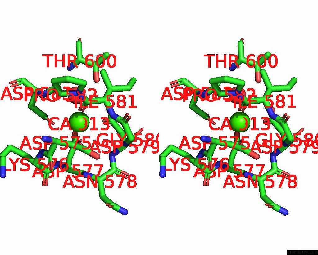
Calcium binding site 1 out of 3 in 8jbo
Go back to
Calcium binding site 1 out
of 3 in the Crystal Structure of TXGH116 From Thermoanaerobacterium Xylanolyticum with Isofagomine
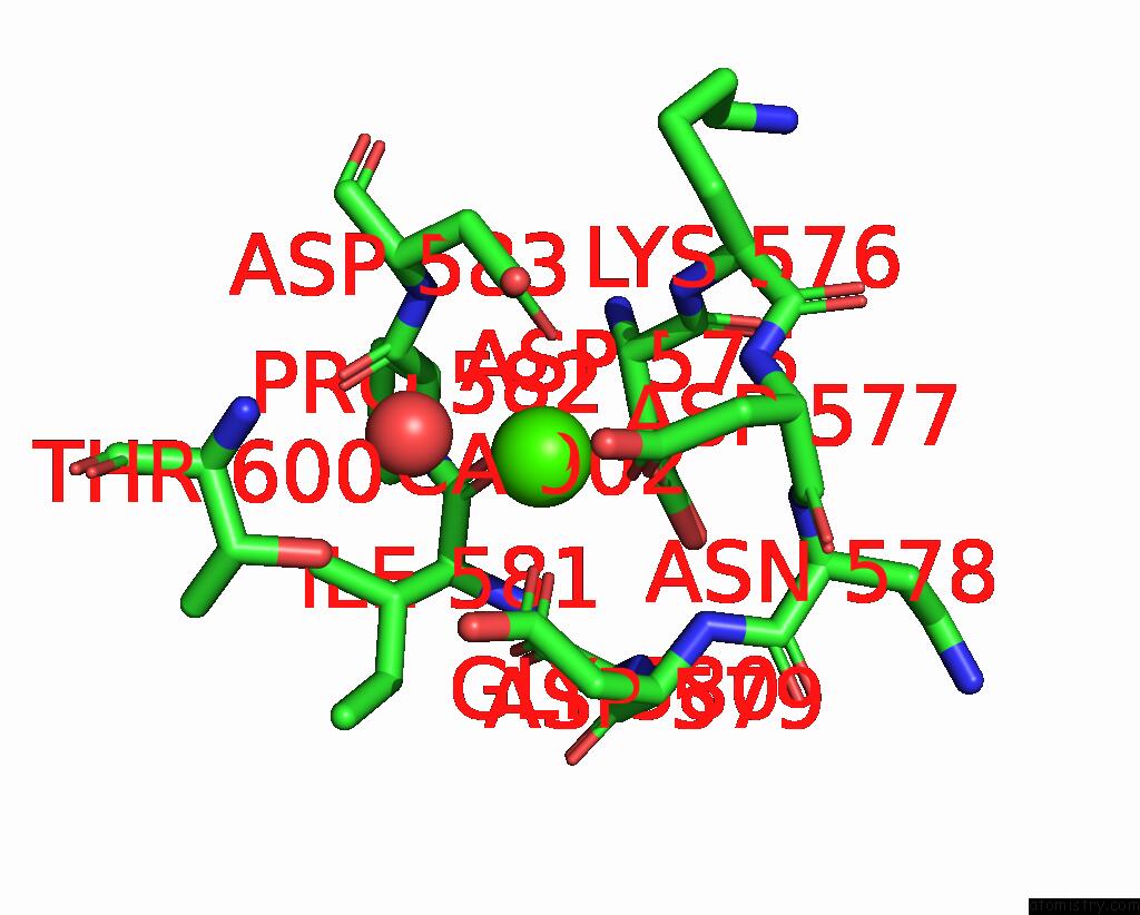
Mono view
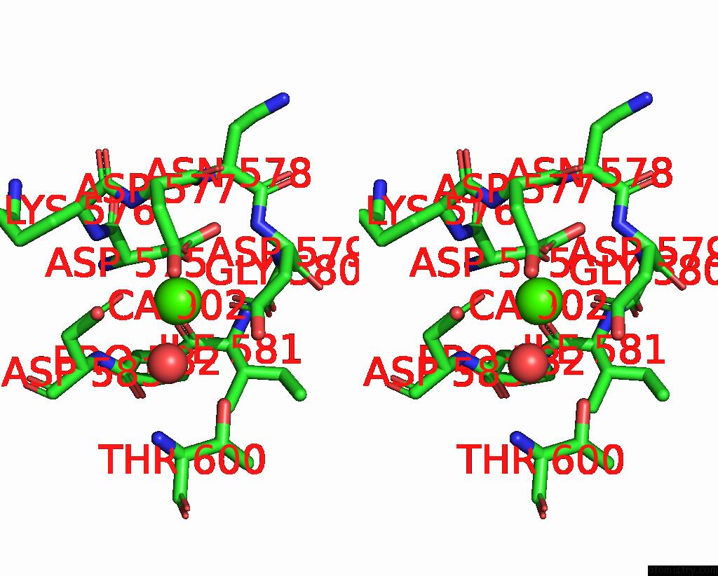
Stereo pair view
Mono view
Stereo pair view
A full contact list of Calcium with other atoms in the Ca binding
site number 1 of Crystal Structure of TXGH116 From Thermoanaerobacterium Xylanolyticum with Isofagomine within 5.0Å range:
|
Calcium binding site 2 out of 3 in 8jbo
Go back to
Calcium binding site 2 out
of 3 in the Crystal Structure of TXGH116 From Thermoanaerobacterium Xylanolyticum with Isofagomine
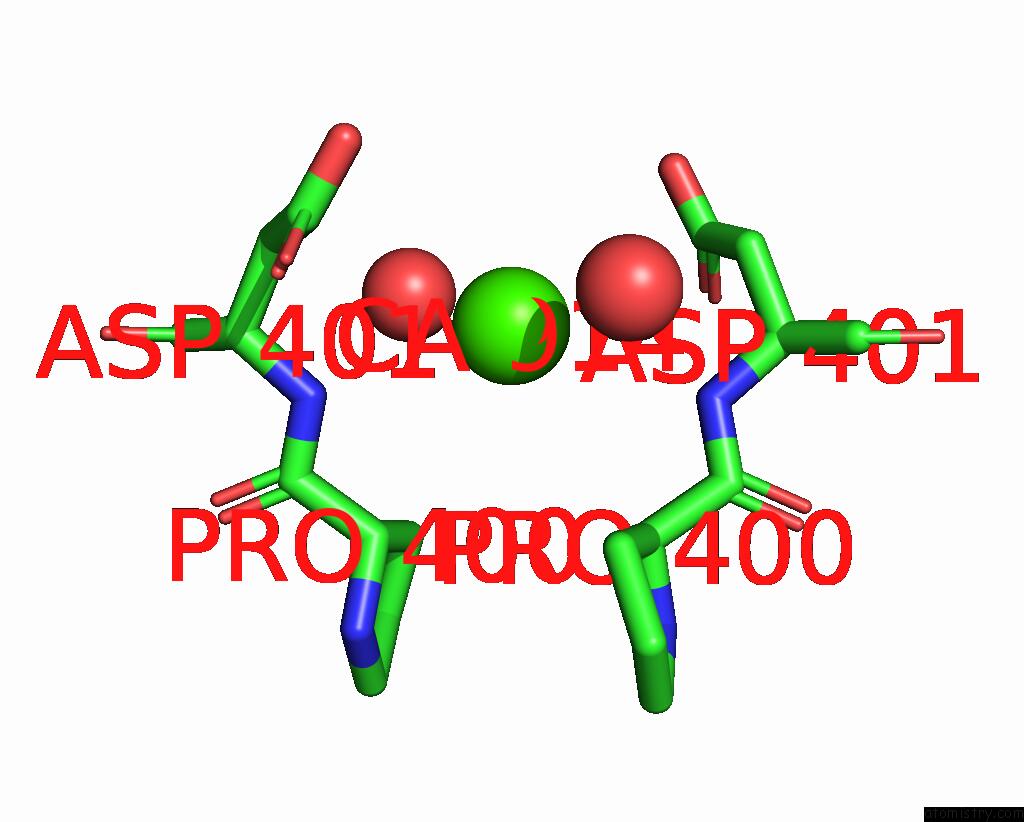
Mono view
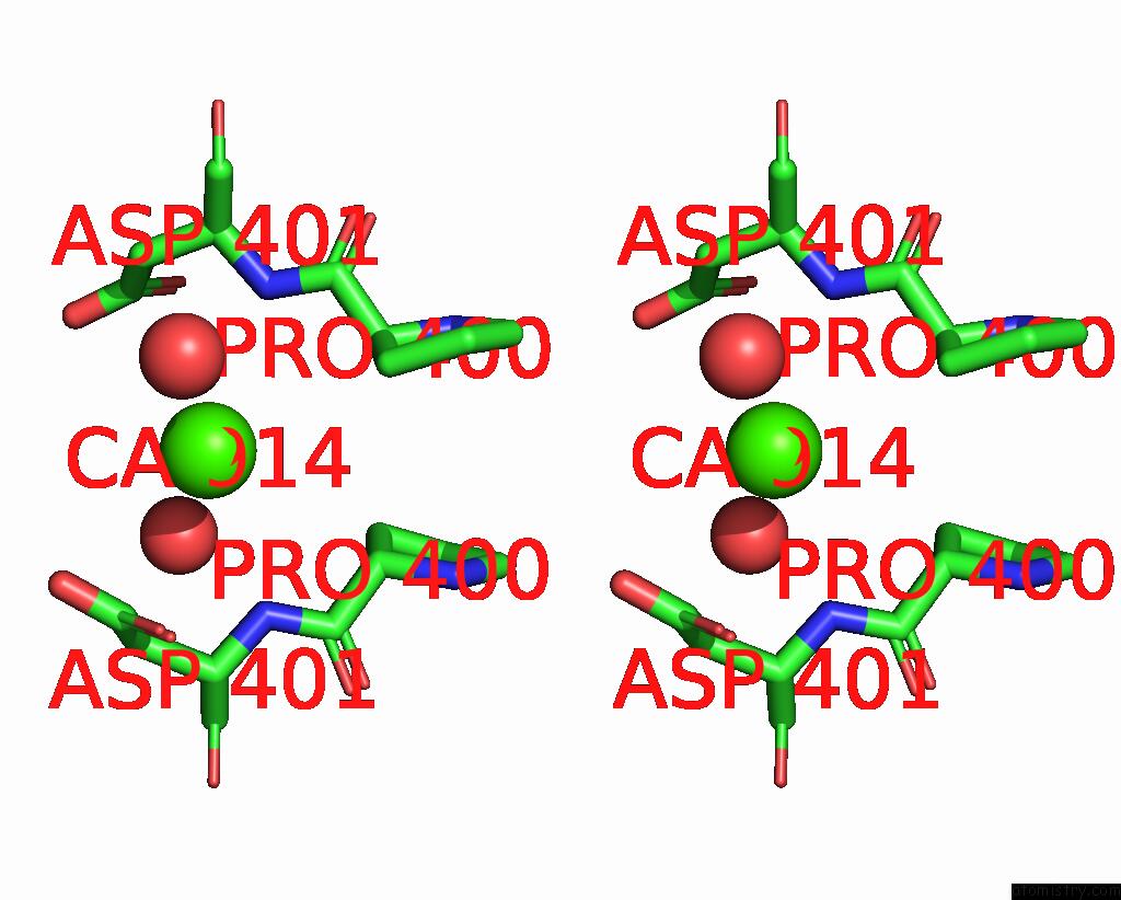
Stereo pair view
Mono view
Stereo pair view
A full contact list of Calcium with other atoms in the Ca binding
site number 2 of Crystal Structure of TXGH116 From Thermoanaerobacterium Xylanolyticum with Isofagomine within 5.0Å range:
|
Calcium binding site 3 out of 3 in 8jbo
Go back to
Calcium binding site 3 out
of 3 in the Crystal Structure of TXGH116 From Thermoanaerobacterium Xylanolyticum with Isofagomine

Mono view
Stereo pair view
Mono view
Stereo pair view
A full contact list of Calcium with other atoms in the Ca binding
site number 3 of Crystal Structure of TXGH116 From Thermoanaerobacterium Xylanolyticum with Isofagomine within 5.0Å range:
|
Reference:
W.Meelua,
N.Thinkumrob,
P.Saparpakorn,
S.Pengthaisong,
S.Hannongbua,
J.R.Ketudat Cairns,
J.Jitonnom.
Structural Basis For Inhibition of A GH116 Beta-Glucosidase and Its Missense Mutants By GBA2 Inhibitors: Crystallographic and Quantum Chemical Study. Chem.Biol.Interact. V. 384 10717 2023.
ISSN: ISSN 0009-2797
PubMed: 37726065
DOI: 10.1016/J.CBI.2023.110717
Page generated: Fri Jul 19 09:57:08 2024
ISSN: ISSN 0009-2797
PubMed: 37726065
DOI: 10.1016/J.CBI.2023.110717
Last articles
Zn in 9JYWZn in 9IR4
Zn in 9IR3
Zn in 9GMX
Zn in 9GMW
Zn in 9JEJ
Zn in 9ERF
Zn in 9ERE
Zn in 9EGV
Zn in 9EGW