Calcium »
PDB 8olc-8q29 »
8owm »
Calcium in PDB 8owm: Crystal Structure of Glutamate Dehydrogenase 2 From Arabidopsis Thaliana Binding Ca, Nad and 2,2-Dihydroxyglutarate
Enzymatic activity of Crystal Structure of Glutamate Dehydrogenase 2 From Arabidopsis Thaliana Binding Ca, Nad and 2,2-Dihydroxyglutarate
All present enzymatic activity of Crystal Structure of Glutamate Dehydrogenase 2 From Arabidopsis Thaliana Binding Ca, Nad and 2,2-Dihydroxyglutarate:
1.4.1.3;
1.4.1.3;
Protein crystallography data
The structure of Crystal Structure of Glutamate Dehydrogenase 2 From Arabidopsis Thaliana Binding Ca, Nad and 2,2-Dihydroxyglutarate, PDB code: 8owm
was solved by
M.Grzechowiak,
M.Ruszkowski,
with X-Ray Crystallography technique. A brief refinement statistics is given in the table below:
| Resolution Low / High (Å) | 65.70 / 1.70 |
| Space group | P 1 |
| Cell size a, b, c (Å), α, β, γ (°) | 95.544, 95.629, 95.841, 90.42, 93.59, 117.78 |
| R / Rfree (%) | 14.4 / 17.2 |
Other elements in 8owm:
The structure of Crystal Structure of Glutamate Dehydrogenase 2 From Arabidopsis Thaliana Binding Ca, Nad and 2,2-Dihydroxyglutarate also contains other interesting chemical elements:
| Sodium | (Na) | 3 atoms |
Calcium Binding Sites:
The binding sites of Calcium atom in the Crystal Structure of Glutamate Dehydrogenase 2 From Arabidopsis Thaliana Binding Ca, Nad and 2,2-Dihydroxyglutarate
(pdb code 8owm). This binding sites where shown within
5.0 Angstroms radius around Calcium atom.
In total 6 binding sites of Calcium where determined in the Crystal Structure of Glutamate Dehydrogenase 2 From Arabidopsis Thaliana Binding Ca, Nad and 2,2-Dihydroxyglutarate, PDB code: 8owm:
Jump to Calcium binding site number: 1; 2; 3; 4; 5; 6;
In total 6 binding sites of Calcium where determined in the Crystal Structure of Glutamate Dehydrogenase 2 From Arabidopsis Thaliana Binding Ca, Nad and 2,2-Dihydroxyglutarate, PDB code: 8owm:
Jump to Calcium binding site number: 1; 2; 3; 4; 5; 6;
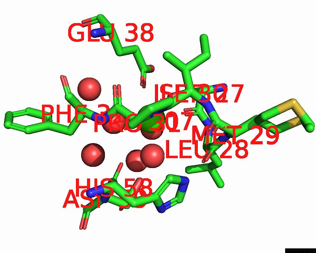
Calcium binding site 1 out of 6 in 8owm
Go back to
Calcium binding site 1 out
of 6 in the Crystal Structure of Glutamate Dehydrogenase 2 From Arabidopsis Thaliana Binding Ca, Nad and 2,2-Dihydroxyglutarate

Mono view
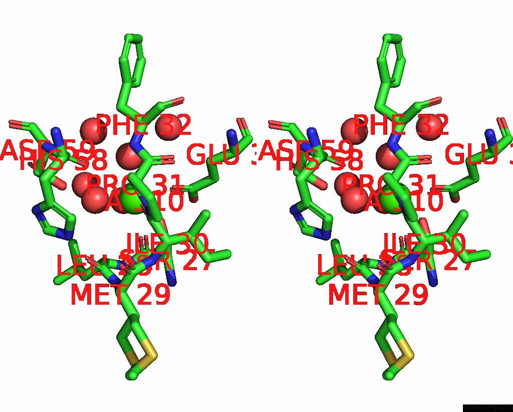
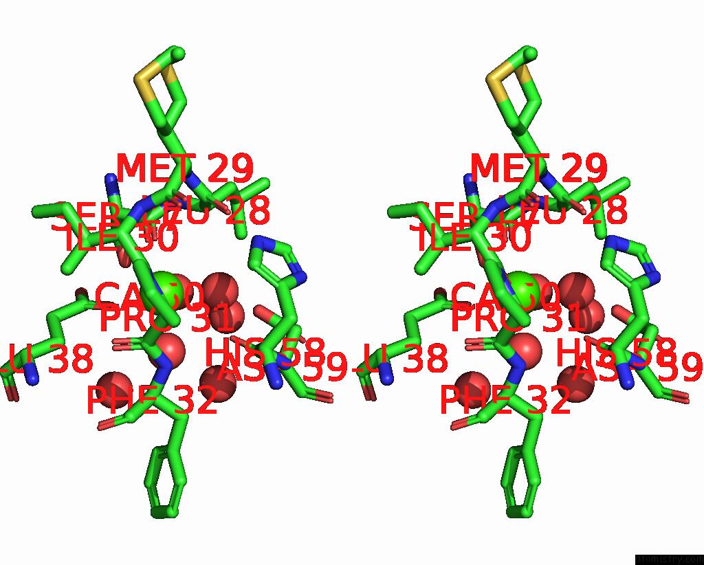
Stereo pair view
Mono view
Stereo pair view
A full contact list of Calcium with other atoms in the Ca binding
site number 1 of Crystal Structure of Glutamate Dehydrogenase 2 From Arabidopsis Thaliana Binding Ca, Nad and 2,2-Dihydroxyglutarate within 5.0Å range:
|
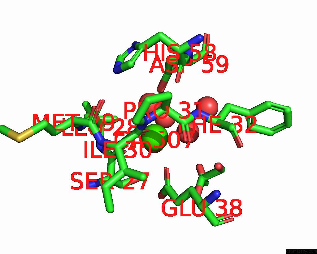
Calcium binding site 2 out of 6 in 8owm
Go back to
Calcium binding site 2 out
of 6 in the Crystal Structure of Glutamate Dehydrogenase 2 From Arabidopsis Thaliana Binding Ca, Nad and 2,2-Dihydroxyglutarate

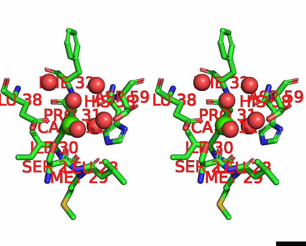
Mono view
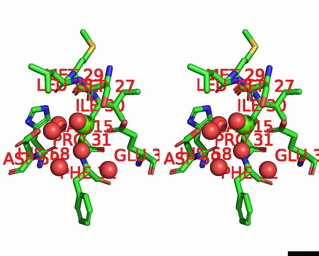
Stereo pair view
Mono view
Stereo pair view
A full contact list of Calcium with other atoms in the Ca binding
site number 2 of Crystal Structure of Glutamate Dehydrogenase 2 From Arabidopsis Thaliana Binding Ca, Nad and 2,2-Dihydroxyglutarate within 5.0Å range:
|
Calcium binding site 3 out of 6 in 8owm
Go back to
Calcium binding site 3 out
of 6 in the Crystal Structure of Glutamate Dehydrogenase 2 From Arabidopsis Thaliana Binding Ca, Nad and 2,2-Dihydroxyglutarate
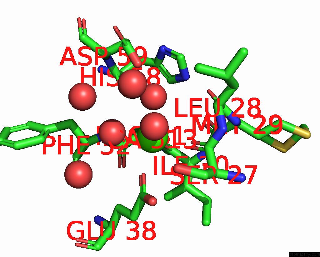
Mono view
Stereo pair view
Mono view
Stereo pair view
A full contact list of Calcium with other atoms in the Ca binding
site number 3 of Crystal Structure of Glutamate Dehydrogenase 2 From Arabidopsis Thaliana Binding Ca, Nad and 2,2-Dihydroxyglutarate within 5.0Å range:
|
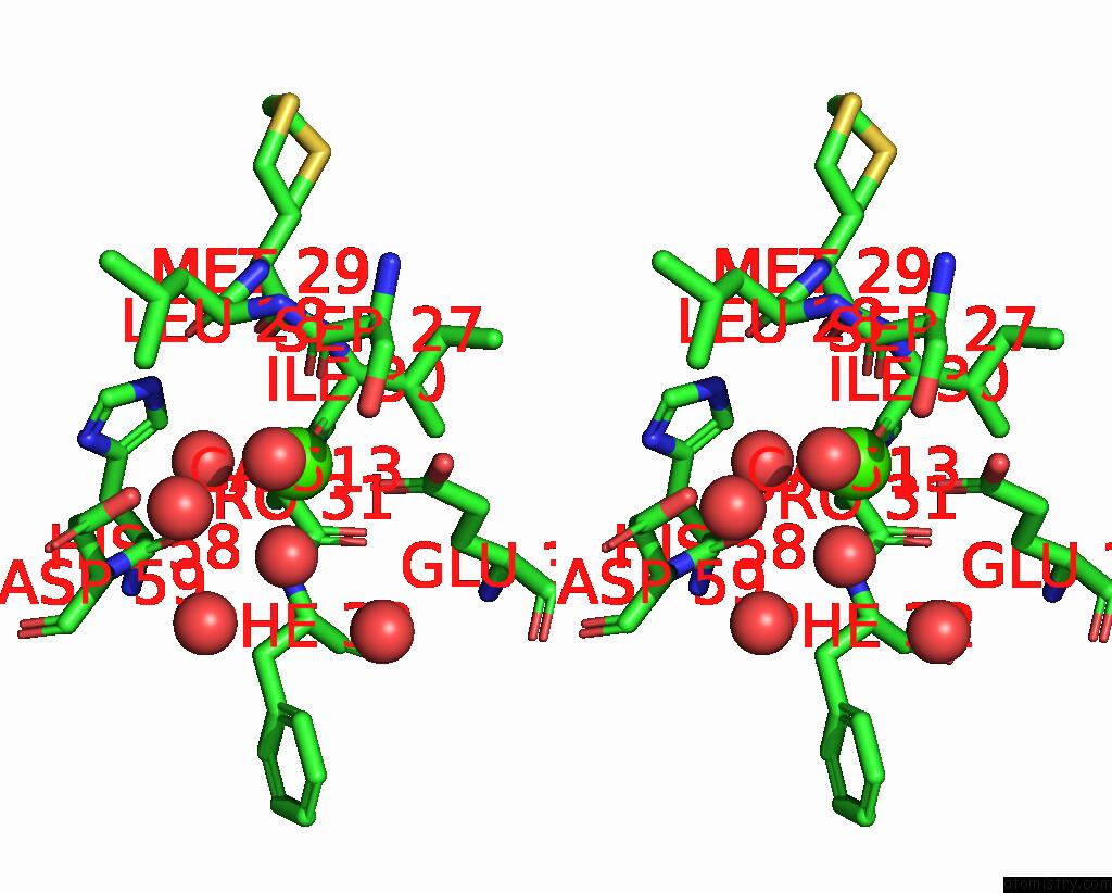
Calcium binding site 4 out of 6 in 8owm
Go back to
Calcium binding site 4 out
of 6 in the Crystal Structure of Glutamate Dehydrogenase 2 From Arabidopsis Thaliana Binding Ca, Nad and 2,2-Dihydroxyglutarate
Mono view
Stereo pair view
Mono view
Stereo pair view
A full contact list of Calcium with other atoms in the Ca binding
site number 4 of Crystal Structure of Glutamate Dehydrogenase 2 From Arabidopsis Thaliana Binding Ca, Nad and 2,2-Dihydroxyglutarate within 5.0Å range:
|
Calcium binding site 5 out of 6 in 8owm
Go back to
Calcium binding site 5 out
of 6 in the Crystal Structure of Glutamate Dehydrogenase 2 From Arabidopsis Thaliana Binding Ca, Nad and 2,2-Dihydroxyglutarate

Mono view
Stereo pair view
Mono view
Stereo pair view
A full contact list of Calcium with other atoms in the Ca binding
site number 5 of Crystal Structure of Glutamate Dehydrogenase 2 From Arabidopsis Thaliana Binding Ca, Nad and 2,2-Dihydroxyglutarate within 5.0Å range:
|
Calcium binding site 6 out of 6 in 8owm
Go back to
Calcium binding site 6 out
of 6 in the Crystal Structure of Glutamate Dehydrogenase 2 From Arabidopsis Thaliana Binding Ca, Nad and 2,2-Dihydroxyglutarate
Mono view

Stereo pair view
Mono view
Stereo pair view
A full contact list of Calcium with other atoms in the Ca binding
site number 6 of Crystal Structure of Glutamate Dehydrogenase 2 From Arabidopsis Thaliana Binding Ca, Nad and 2,2-Dihydroxyglutarate within 5.0Å range:
|
Reference:
M.Grzechowiak,
J.Sliwiak,
M.Jaskolski,
M.Ruszkowski.
Structural and Functional Studies of Arabidopsis Thaliana Glutamate Dehydrogenase Isoform 2 Demonstrate Enzyme Dynamics and Identify Its Calcium Binding Site. Plant Physiol Biochem. V. 201 07895 2023.
ISSN: ESSN 1873-2690
PubMed: 37478728
DOI: 10.1016/J.PLAPHY.2023.107895
Page generated: Thu Jul 10 06:22:05 2025
ISSN: ESSN 1873-2690
PubMed: 37478728
DOI: 10.1016/J.PLAPHY.2023.107895
Last articles
F in 7MYYF in 7N13
F in 7MYU
F in 7MYR
F in 7MYO
F in 7MXN
F in 7MXG
F in 7MXH
F in 7MX7
F in 7MVS