Calcium »
PDB 8olc-8q29 »
8pjx »
Calcium in PDB 8pjx: Crystal Structure of the Computationally Designed SAKE6FR Protein
Protein crystallography data
The structure of Crystal Structure of the Computationally Designed SAKE6FR Protein, PDB code: 8pjx
was solved by
S.M.L.Wouters,
with X-Ray Crystallography technique. A brief refinement statistics is given in the table below:
| Resolution Low / High (Å) | 48.11 / 1.70 |
| Space group | P 43 21 2 |
| Cell size a, b, c (Å), α, β, γ (°) | 68.032, 68.032, 117.857, 90, 90, 90 |
| R / Rfree (%) | 18.2 / 18.5 |
Calcium Binding Sites:
The binding sites of Calcium atom in the Crystal Structure of the Computationally Designed SAKE6FR Protein
(pdb code 8pjx). This binding sites where shown within
5.0 Angstroms radius around Calcium atom.
In total 3 binding sites of Calcium where determined in the Crystal Structure of the Computationally Designed SAKE6FR Protein, PDB code: 8pjx:
Jump to Calcium binding site number: 1; 2; 3;
In total 3 binding sites of Calcium where determined in the Crystal Structure of the Computationally Designed SAKE6FR Protein, PDB code: 8pjx:
Jump to Calcium binding site number: 1; 2; 3;
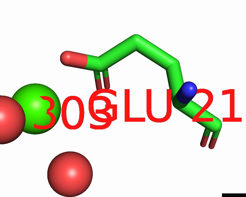
Calcium binding site 1 out of 3 in 8pjx
Go back to
Calcium binding site 1 out
of 3 in the Crystal Structure of the Computationally Designed SAKE6FR Protein
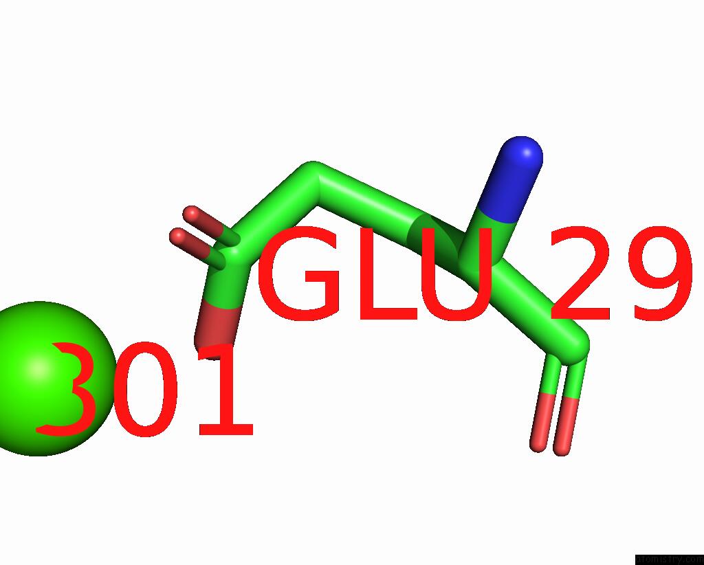
Mono view
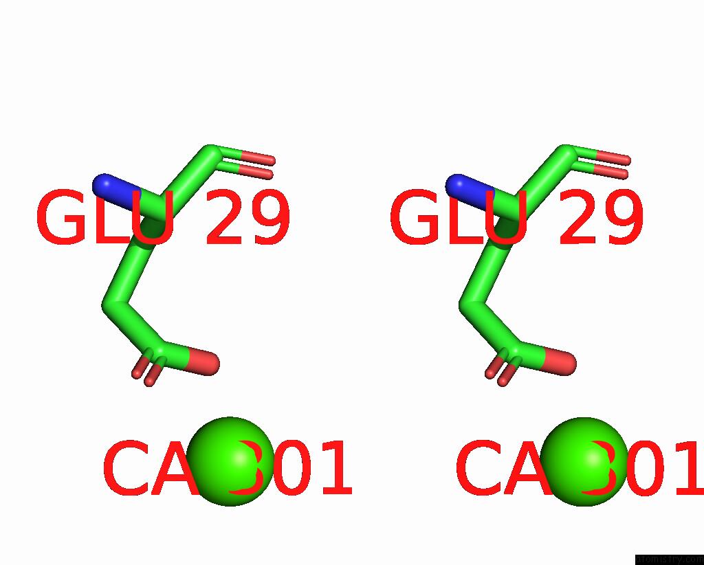
Stereo pair view
Mono view
Stereo pair view
A full contact list of Calcium with other atoms in the Ca binding
site number 1 of Crystal Structure of the Computationally Designed SAKE6FR Protein within 5.0Å range:
|
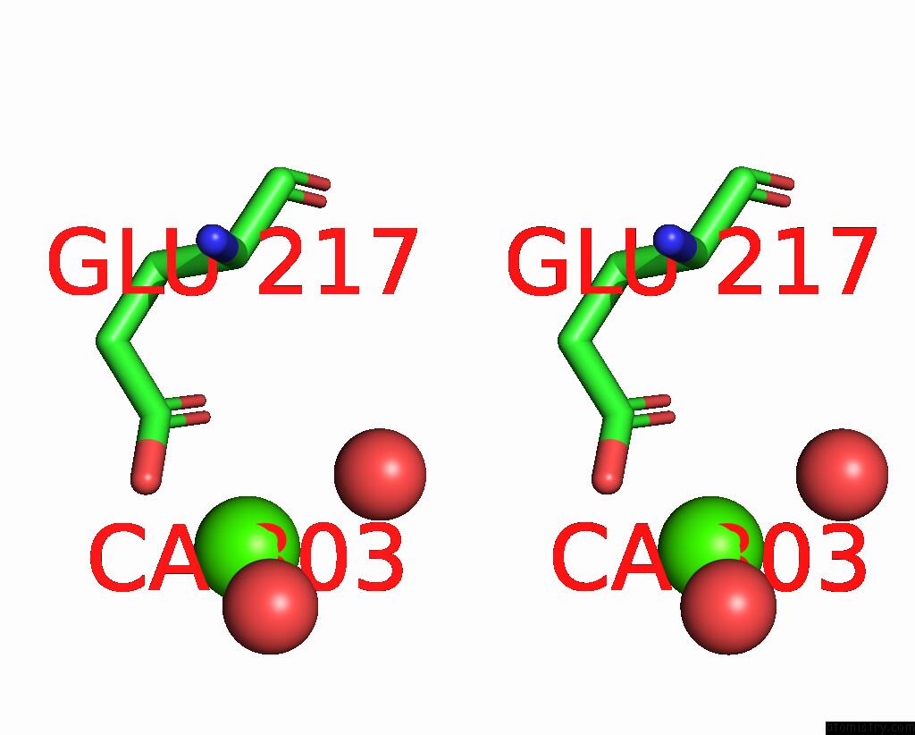
Calcium binding site 2 out of 3 in 8pjx
Go back to
Calcium binding site 2 out
of 3 in the Crystal Structure of the Computationally Designed SAKE6FR Protein
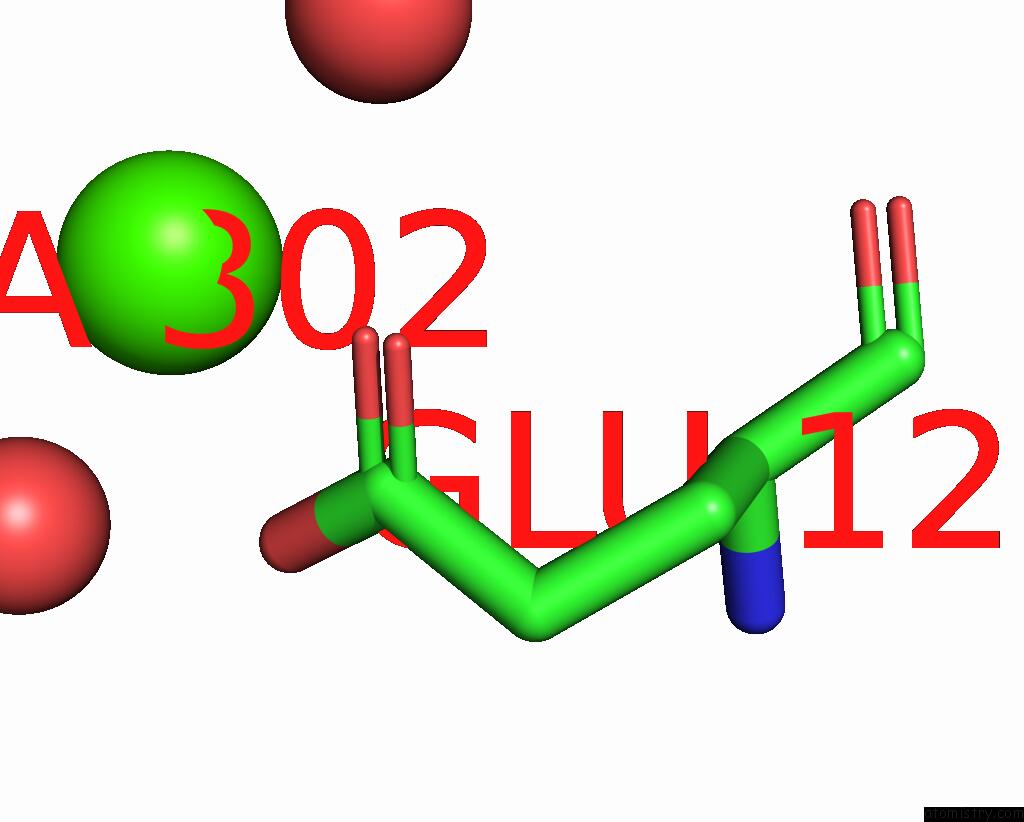
Mono view
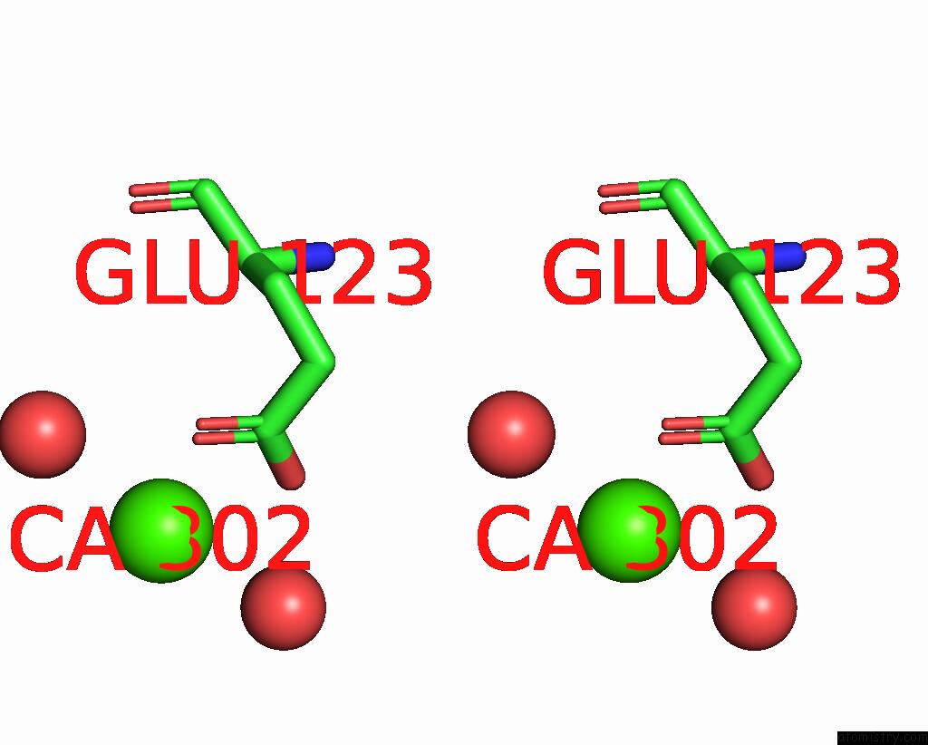
Stereo pair view
Mono view
Stereo pair view
A full contact list of Calcium with other atoms in the Ca binding
site number 2 of Crystal Structure of the Computationally Designed SAKE6FR Protein within 5.0Å range:
|
Calcium binding site 3 out of 3 in 8pjx
Go back to
Calcium binding site 3 out
of 3 in the Crystal Structure of the Computationally Designed SAKE6FR Protein
Mono view
Stereo pair view
Mono view
Stereo pair view
A full contact list of Calcium with other atoms in the Ca binding
site number 3 of Crystal Structure of the Computationally Designed SAKE6FR Protein within 5.0Å range:
|
Reference:
S.M.L.Wouters,
H.Noguchi,
A.R.D.Voet.
Computational Design of the Sake Scaffold Proteins To Be Published.
Page generated: Thu Jul 10 06:27:50 2025
Last articles
Cl in 5RAQCl in 5RAP
Cl in 5RAO
Cl in 5RAN
Cl in 5RAJ
Cl in 5RAM
Cl in 5RAL
Cl in 5RAK
Cl in 5RAF
Cl in 5RAG