Calcium »
PDB 8uws-8vh0 »
8v0i »
Calcium in PDB 8v0i: The Structure of the Native Cardiac Thin Filament Troponin Core in CA2+-Bound Fully Activated State 2 From the Lower Strand
Other elements in 8v0i:
The structure of The Structure of the Native Cardiac Thin Filament Troponin Core in CA2+-Bound Fully Activated State 2 From the Lower Strand also contains other interesting chemical elements:
| Magnesium | (Mg) | 2 atoms |
Calcium Binding Sites:
The binding sites of Calcium atom in the The Structure of the Native Cardiac Thin Filament Troponin Core in CA2+-Bound Fully Activated State 2 From the Lower Strand
(pdb code 8v0i). This binding sites where shown within
5.0 Angstroms radius around Calcium atom.
In total 3 binding sites of Calcium where determined in the The Structure of the Native Cardiac Thin Filament Troponin Core in CA2+-Bound Fully Activated State 2 From the Lower Strand, PDB code: 8v0i:
Jump to Calcium binding site number: 1; 2; 3;
In total 3 binding sites of Calcium where determined in the The Structure of the Native Cardiac Thin Filament Troponin Core in CA2+-Bound Fully Activated State 2 From the Lower Strand, PDB code: 8v0i:
Jump to Calcium binding site number: 1; 2; 3;
Calcium binding site 1 out of 3 in 8v0i
Go back to
Calcium binding site 1 out
of 3 in the The Structure of the Native Cardiac Thin Filament Troponin Core in CA2+-Bound Fully Activated State 2 From the Lower Strand
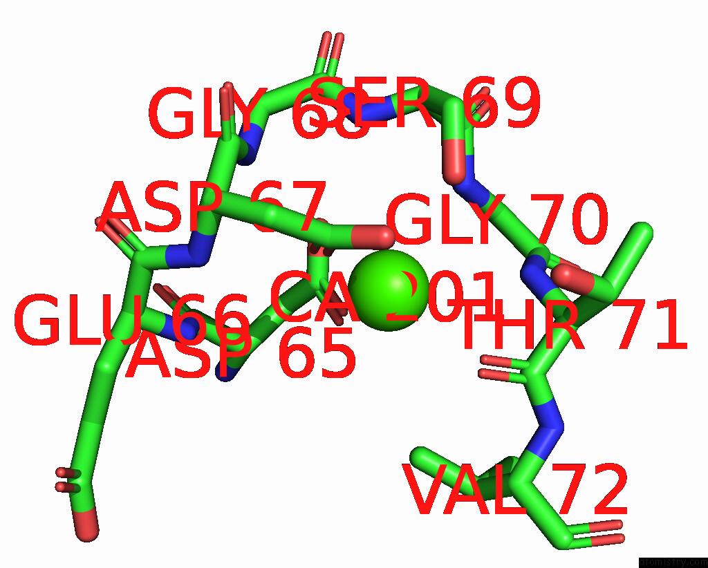
Mono view
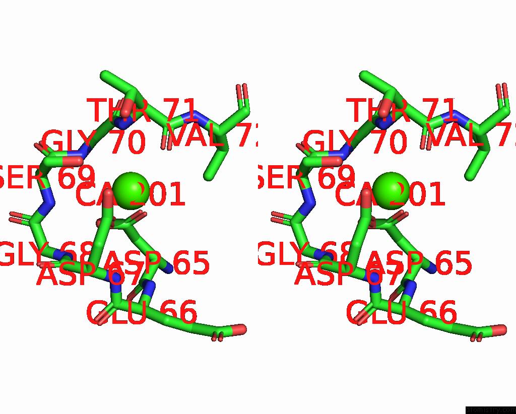
Stereo pair view

Mono view

Stereo pair view
A full contact list of Calcium with other atoms in the Ca binding
site number 1 of The Structure of the Native Cardiac Thin Filament Troponin Core in CA2+-Bound Fully Activated State 2 From the Lower Strand within 5.0Å range:
|
Calcium binding site 2 out of 3 in 8v0i
Go back to
Calcium binding site 2 out
of 3 in the The Structure of the Native Cardiac Thin Filament Troponin Core in CA2+-Bound Fully Activated State 2 From the Lower Strand
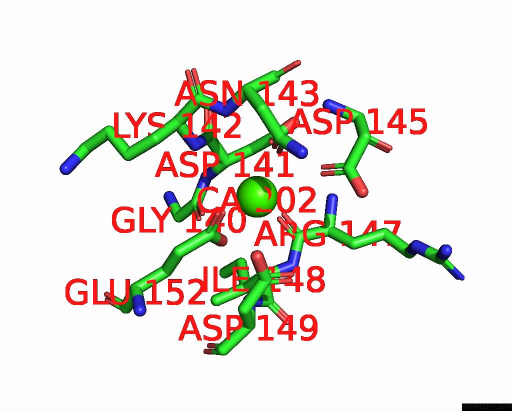
Mono view
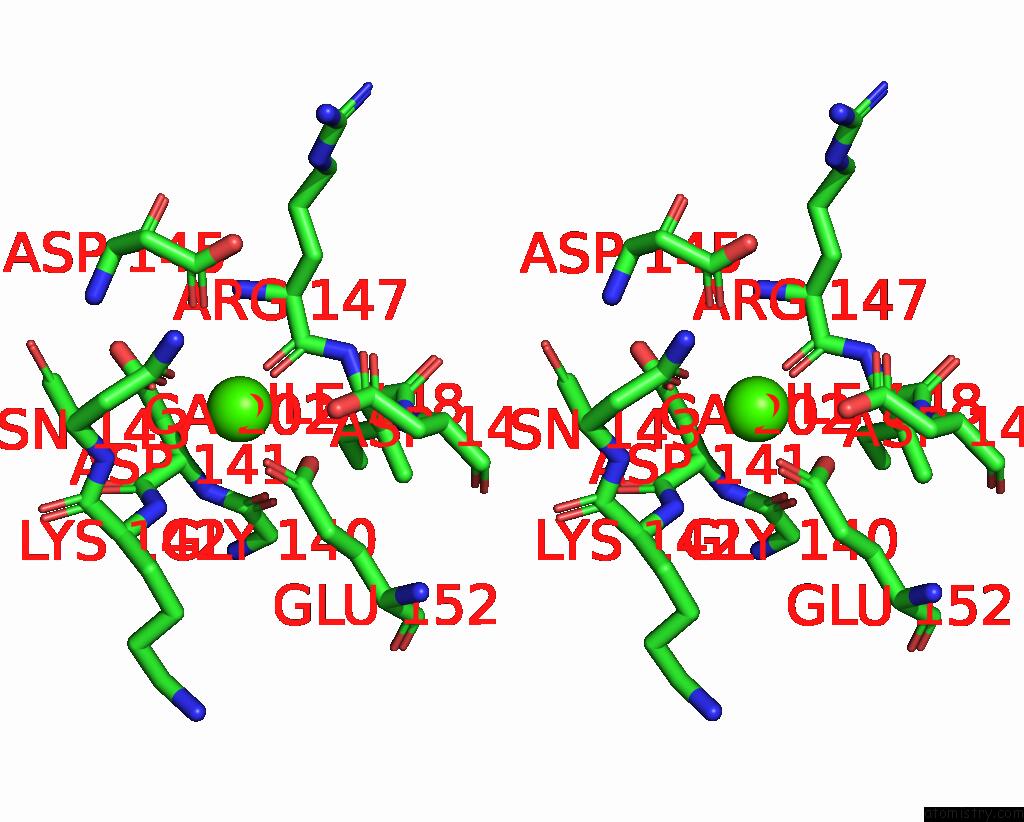
Stereo pair view

Mono view

Stereo pair view
A full contact list of Calcium with other atoms in the Ca binding
site number 2 of The Structure of the Native Cardiac Thin Filament Troponin Core in CA2+-Bound Fully Activated State 2 From the Lower Strand within 5.0Å range:
|
Calcium binding site 3 out of 3 in 8v0i
Go back to
Calcium binding site 3 out
of 3 in the The Structure of the Native Cardiac Thin Filament Troponin Core in CA2+-Bound Fully Activated State 2 From the Lower Strand
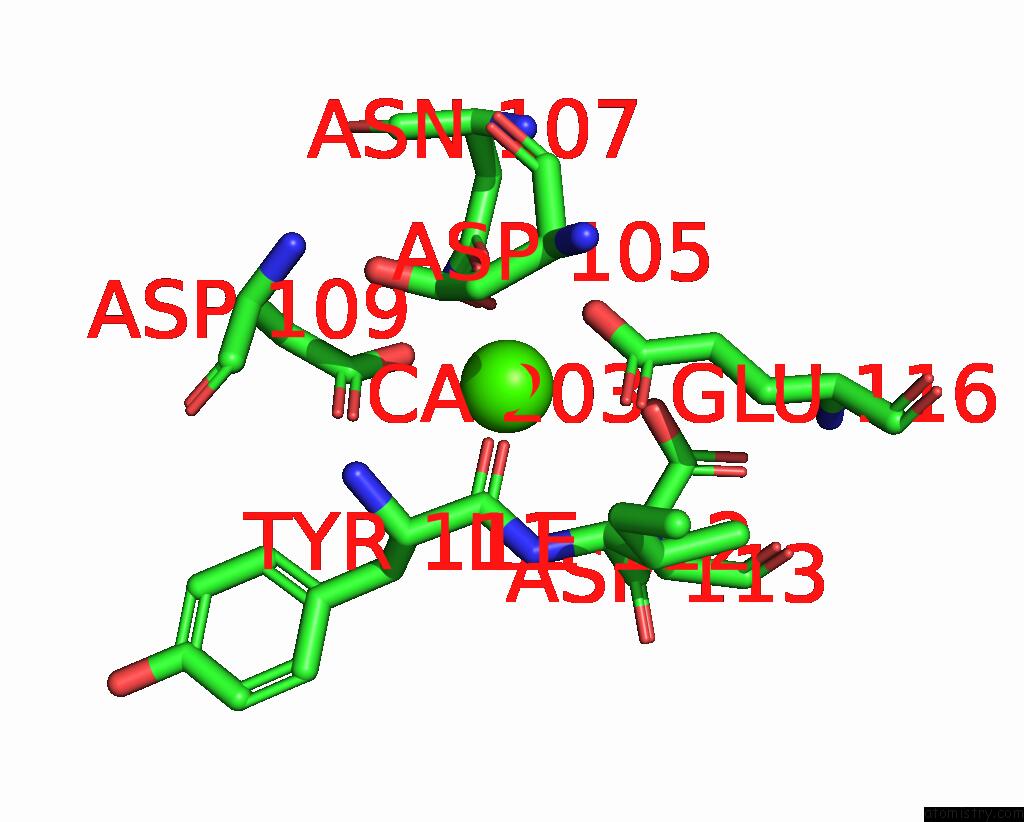
Mono view
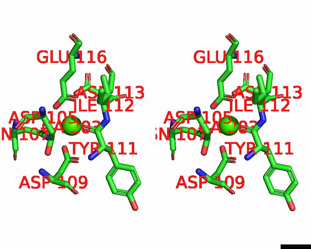
Stereo pair view

Mono view

Stereo pair view
A full contact list of Calcium with other atoms in the Ca binding
site number 3 of The Structure of the Native Cardiac Thin Filament Troponin Core in CA2+-Bound Fully Activated State 2 From the Lower Strand within 5.0Å range:
|
Reference:
C.M.Risi,
B.Belknap,
J.Atherton,
I.Leite Coscarella,
H.D.White,
P.Bryant Chase,
J.R.Pinto,
V.E.Galkin.
Troponin Structural Dynamics in the Native Cardiac Thin Filament Revealed By Cryo Electron Microscopy. J.Mol.Biol. 68498 2024.
ISSN: ESSN 1089-8638
PubMed: 38387550
DOI: 10.1016/J.JMB.2024.168498
Page generated: Thu Jul 10 07:53:53 2025
ISSN: ESSN 1089-8638
PubMed: 38387550
DOI: 10.1016/J.JMB.2024.168498
Last articles
F in 7KZ4F in 7KYV
F in 7KYT
F in 7KYB
F in 7KYC
F in 7KYA
F in 7KY5
F in 7KXW
F in 7KY9
F in 7KWA