Calcium »
PDB 8uws-8vh0 »
8v9o »
Calcium in PDB 8v9o: Imaging Scaffold Engineered to Bind the Therapeutic Protein Target BARD1
Protein crystallography data
The structure of Imaging Scaffold Engineered to Bind the Therapeutic Protein Target BARD1, PDB code: 8v9o
was solved by
M.P.Agdanowski,
R.Castells-Graells,
M.R.Sawaya,
T.O.Yeates,
M.A.Arbing,
with X-Ray Crystallography technique. A brief refinement statistics is given in the table below:
| Resolution Low / High (Å) | 19.99 / 3.81 |
| Space group | I 2 2 2 |
| Cell size a, b, c (Å), α, β, γ (°) | 128.01, 195.57, 228.37, 90, 90, 90 |
| R / Rfree (%) | 19.7 / 23.1 |
Calcium Binding Sites:
The binding sites of Calcium atom in the Imaging Scaffold Engineered to Bind the Therapeutic Protein Target BARD1
(pdb code 8v9o). This binding sites where shown within
5.0 Angstroms radius around Calcium atom.
In total only one binding site of Calcium was determined in the Imaging Scaffold Engineered to Bind the Therapeutic Protein Target BARD1, PDB code: 8v9o:
In total only one binding site of Calcium was determined in the Imaging Scaffold Engineered to Bind the Therapeutic Protein Target BARD1, PDB code: 8v9o:
Calcium binding site 1 out of 1 in 8v9o
Go back to
Calcium binding site 1 out
of 1 in the Imaging Scaffold Engineered to Bind the Therapeutic Protein Target BARD1
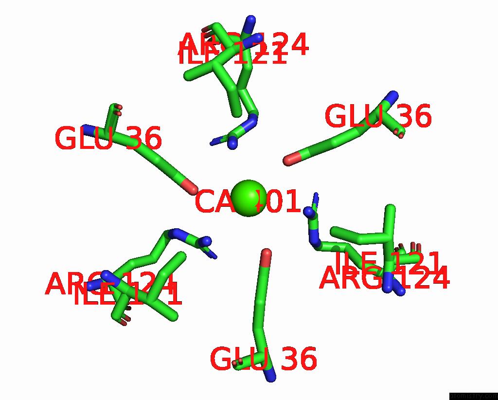
Mono view
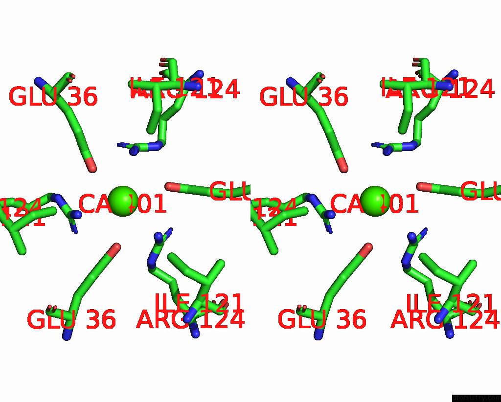
Stereo pair view
Mono view
Stereo pair view
A full contact list of Calcium with other atoms in the Ca binding
site number 1 of Imaging Scaffold Engineered to Bind the Therapeutic Protein Target BARD1 within 5.0Å range:
|
Reference:
M.P.Agdanowski,
R.Castells-Graells,
M.R.Sawaya,
T.O.Yeates,
M.A.Arbing.
X-Ray Structure of A Designed Rigidified Imaging Scaffold Engineered to Bind the Therapeutic Protein Target BARD1 To Be Published.
Page generated: Thu Jul 10 07:58:59 2025
Last articles
Cl in 5I13Cl in 5I1C
Cl in 5I0W
Cl in 5I0V
Cl in 5HZX
Cl in 5I0R
Cl in 5I02
Cl in 5I00
Cl in 5HZ5
Cl in 5HZ8