Calcium »
PDB 8vh8-8wbt »
8vh8 »
Calcium in PDB 8vh8: Crystal Structure of Heparosan Synthase 2 From Pasteurella Multocida at 2.85 A
Protein crystallography data
The structure of Crystal Structure of Heparosan Synthase 2 From Pasteurella Multocida at 2.85 A, PDB code: 8vh8
was solved by
L.C.Pedersen,
J.Liu,
E.Stancanelli,
J.M.Krahn,
with X-Ray Crystallography technique. A brief refinement statistics is given in the table below:
| Resolution Low / High (Å) | 28.76 / 2.85 |
| Space group | P 43 |
| Cell size a, b, c (Å), α, β, γ (°) | 118.57, 118.57, 262.356, 90, 90, 90 |
| R / Rfree (%) | 19.8 / 22.3 |
Other elements in 8vh8:
The structure of Crystal Structure of Heparosan Synthase 2 From Pasteurella Multocida at 2.85 A also contains other interesting chemical elements:
| Sodium | (Na) | 4 atoms |
| Manganese | (Mn) | 8 atoms |
| Chlorine | (Cl) | 18 atoms |
Calcium Binding Sites:
The binding sites of Calcium atom in the Crystal Structure of Heparosan Synthase 2 From Pasteurella Multocida at 2.85 A
(pdb code 8vh8). This binding sites where shown within
5.0 Angstroms radius around Calcium atom.
In total only one binding site of Calcium was determined in the Crystal Structure of Heparosan Synthase 2 From Pasteurella Multocida at 2.85 A, PDB code: 8vh8:
In total only one binding site of Calcium was determined in the Crystal Structure of Heparosan Synthase 2 From Pasteurella Multocida at 2.85 A, PDB code: 8vh8:
Calcium binding site 1 out of 1 in 8vh8
Go back to
Calcium binding site 1 out
of 1 in the Crystal Structure of Heparosan Synthase 2 From Pasteurella Multocida at 2.85 A
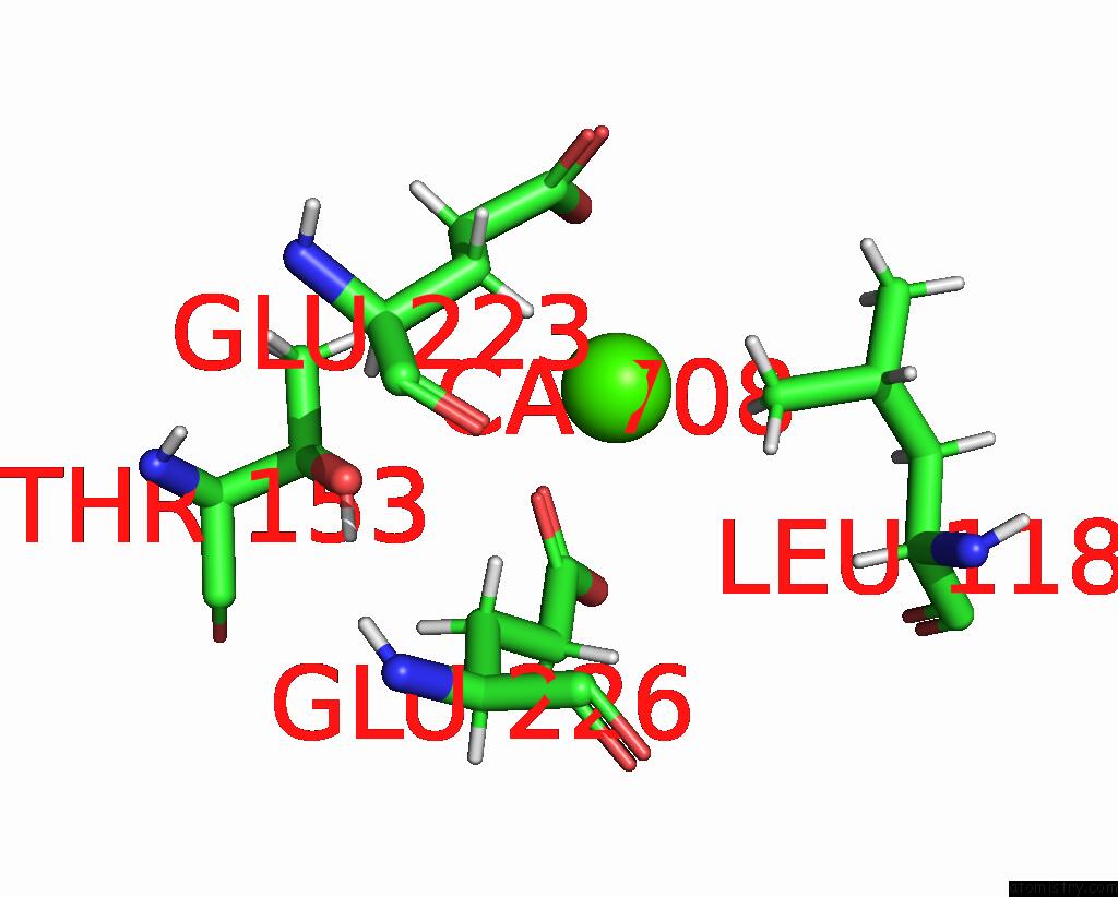
Mono view
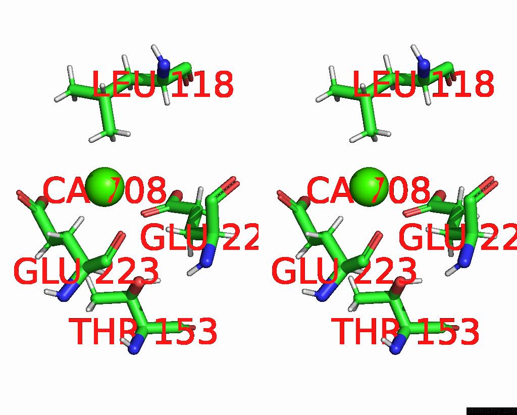
Stereo pair view
Mono view
Stereo pair view
A full contact list of Calcium with other atoms in the Ca binding
site number 1 of Crystal Structure of Heparosan Synthase 2 From Pasteurella Multocida at 2.85 A within 5.0Å range:
|
Reference:
E.Stancanelli,
J.A.Krahn,
E.Viverette,
R.Dutcher,
V.Pagadala,
M.J.Borgnia,
J.Liu,
L.C.Pedersen.
Structural and Functional Analysis of Heparosan Synthase 2 From Pasteurella Multocida to Improve the Synthesis of Heparin Acs Catalysis V. 14 6577 2024.
ISSN: ESSN 2155-5435
DOI: 10.1021/ACSCATAL.4C00677
Page generated: Thu Jul 10 08:00:28 2025
ISSN: ESSN 2155-5435
DOI: 10.1021/ACSCATAL.4C00677
Last articles
Fe in 2YXOFe in 2YRS
Fe in 2YXC
Fe in 2YNM
Fe in 2YVJ
Fe in 2YP1
Fe in 2YU2
Fe in 2YU1
Fe in 2YQB
Fe in 2YOO