Calcium »
PDB 8vh8-8wbt »
8vha »
Calcium in PDB 8vha: Crystal Structure of Human IDH1 R132Q in Complex with Nadph and Alpha- Ketoglutarate
Enzymatic activity of Crystal Structure of Human IDH1 R132Q in Complex with Nadph and Alpha- Ketoglutarate
All present enzymatic activity of Crystal Structure of Human IDH1 R132Q in Complex with Nadph and Alpha- Ketoglutarate:
1.1.1.42;
1.1.1.42;
Protein crystallography data
The structure of Crystal Structure of Human IDH1 R132Q in Complex with Nadph and Alpha- Ketoglutarate, PDB code: 8vha
was solved by
M.Mealka,
C.D.Sohl,
T.Huxford,
with X-Ray Crystallography technique. A brief refinement statistics is given in the table below:
| Resolution Low / High (Å) | 39.15 / 2.28 |
| Space group | P 1 21 1 |
| Cell size a, b, c (Å), α, β, γ (°) | 83.821, 104.86, 107.711, 90, 98.19, 90 |
| R / Rfree (%) | 16.7 / 22.2 |
Calcium Binding Sites:
The binding sites of Calcium atom in the Crystal Structure of Human IDH1 R132Q in Complex with Nadph and Alpha- Ketoglutarate
(pdb code 8vha). This binding sites where shown within
5.0 Angstroms radius around Calcium atom.
In total 4 binding sites of Calcium where determined in the Crystal Structure of Human IDH1 R132Q in Complex with Nadph and Alpha- Ketoglutarate, PDB code: 8vha:
Jump to Calcium binding site number: 1; 2; 3; 4;
In total 4 binding sites of Calcium where determined in the Crystal Structure of Human IDH1 R132Q in Complex with Nadph and Alpha- Ketoglutarate, PDB code: 8vha:
Jump to Calcium binding site number: 1; 2; 3; 4;
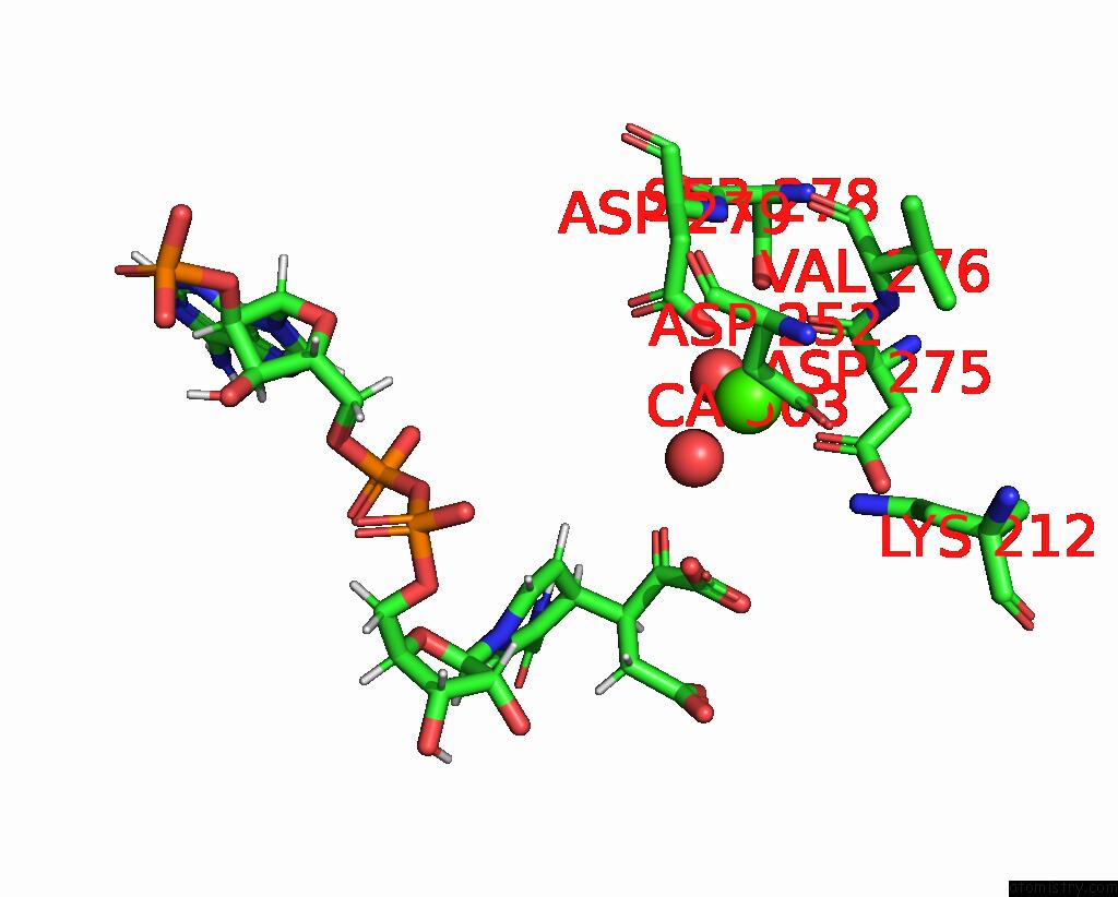
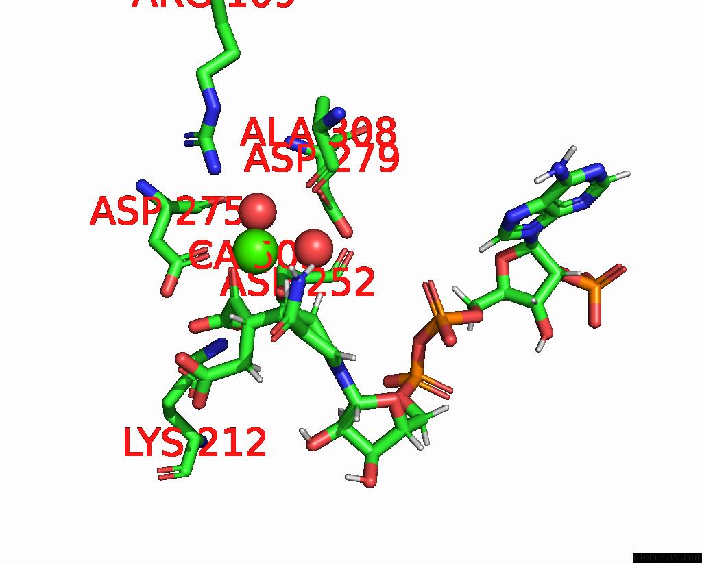
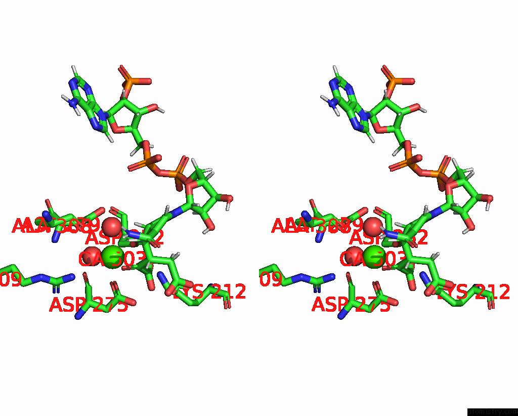
Calcium binding site 1 out of 4 in 8vha
Go back to
Calcium binding site 1 out
of 4 in the Crystal Structure of Human IDH1 R132Q in Complex with Nadph and Alpha- Ketoglutarate
Mono view
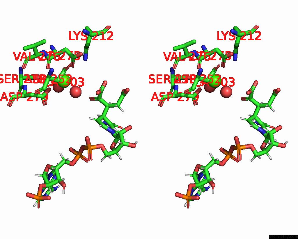
Stereo pair view
Mono view
Stereo pair view
A full contact list of Calcium with other atoms in the Ca binding
site number 1 of Crystal Structure of Human IDH1 R132Q in Complex with Nadph and Alpha- Ketoglutarate within 5.0Å range:
|
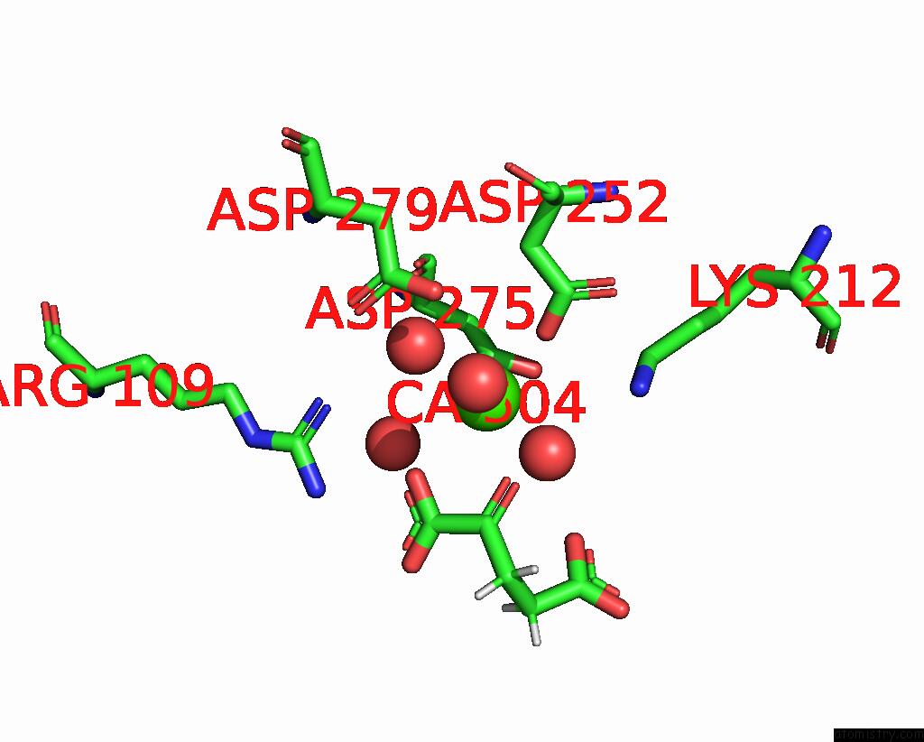
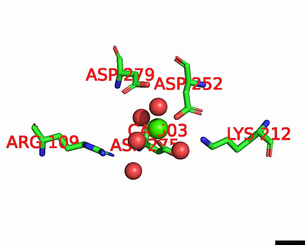
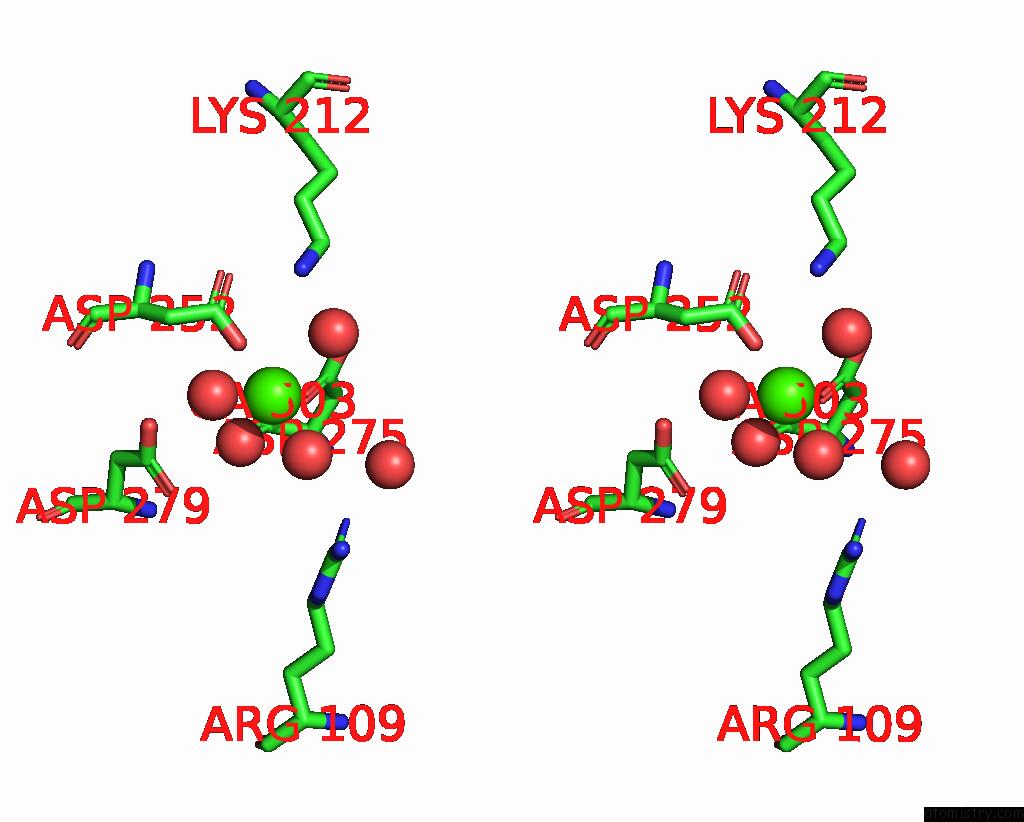
Calcium binding site 2 out of 4 in 8vha
Go back to
Calcium binding site 2 out
of 4 in the Crystal Structure of Human IDH1 R132Q in Complex with Nadph and Alpha- Ketoglutarate
Mono view
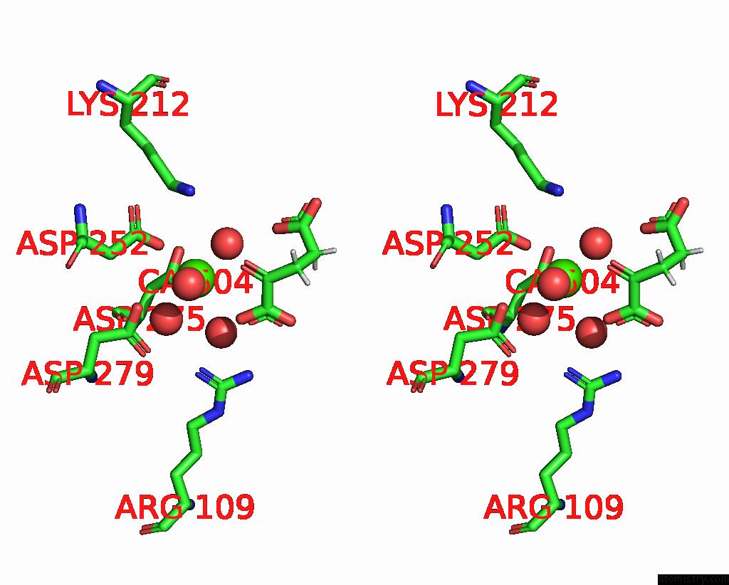
Stereo pair view
Mono view
Stereo pair view
A full contact list of Calcium with other atoms in the Ca binding
site number 2 of Crystal Structure of Human IDH1 R132Q in Complex with Nadph and Alpha- Ketoglutarate within 5.0Å range:
|
Calcium binding site 3 out of 4 in 8vha
Go back to
Calcium binding site 3 out
of 4 in the Crystal Structure of Human IDH1 R132Q in Complex with Nadph and Alpha- Ketoglutarate
Mono view
Stereo pair view
Mono view
Stereo pair view
A full contact list of Calcium with other atoms in the Ca binding
site number 3 of Crystal Structure of Human IDH1 R132Q in Complex with Nadph and Alpha- Ketoglutarate within 5.0Å range:
|
Calcium binding site 4 out of 4 in 8vha
Go back to
Calcium binding site 4 out
of 4 in the Crystal Structure of Human IDH1 R132Q in Complex with Nadph and Alpha- Ketoglutarate
Mono view
Stereo pair view
Mono view
Stereo pair view
A full contact list of Calcium with other atoms in the Ca binding
site number 4 of Crystal Structure of Human IDH1 R132Q in Complex with Nadph and Alpha- Ketoglutarate within 5.0Å range:
|
Reference:
M.Mealka,
C.D.Sohl,
T.Huxford.
Active Site Remodeling in Tumor-Relevant IDH1 Mutants Drive Distinct Kinetic Features and Possible Resistance Mechanisms To Be Published.
Page generated: Thu Jul 10 08:00:26 2025
Last articles
Fe in 2YXOFe in 2YRS
Fe in 2YXC
Fe in 2YNM
Fe in 2YVJ
Fe in 2YP1
Fe in 2YU2
Fe in 2YU1
Fe in 2YQB
Fe in 2YOO