Calcium »
PDB 8vh8-8wbt »
8wbt »
Calcium in PDB 8wbt: Crystal Structure of Cis-Epoxysuccinate Hydrolases Klcesh[L] Mutant D48N Complexed with L-Ta
Protein crystallography data
The structure of Crystal Structure of Cis-Epoxysuccinate Hydrolases Klcesh[L] Mutant D48N Complexed with L-Ta, PDB code: 8wbt
was solved by
S.Dong,
J.S.Xuan,
Y.G.Feng,
Q.Cui,
with X-Ray Crystallography technique. A brief refinement statistics is given in the table below:
| Resolution Low / High (Å) | 30.29 / 2.05 |
| Space group | P 21 21 2 |
| Cell size a, b, c (Å), α, β, γ (°) | 119.62, 93.15, 101.64, 90, 90, 90 |
| R / Rfree (%) | 16.8 / 20.7 |
Calcium Binding Sites:
The binding sites of Calcium atom in the Crystal Structure of Cis-Epoxysuccinate Hydrolases Klcesh[L] Mutant D48N Complexed with L-Ta
(pdb code 8wbt). This binding sites where shown within
5.0 Angstroms radius around Calcium atom.
In total 2 binding sites of Calcium where determined in the Crystal Structure of Cis-Epoxysuccinate Hydrolases Klcesh[L] Mutant D48N Complexed with L-Ta, PDB code: 8wbt:
Jump to Calcium binding site number: 1; 2;
In total 2 binding sites of Calcium where determined in the Crystal Structure of Cis-Epoxysuccinate Hydrolases Klcesh[L] Mutant D48N Complexed with L-Ta, PDB code: 8wbt:
Jump to Calcium binding site number: 1; 2;
Calcium binding site 1 out of 2 in 8wbt
Go back to
Calcium binding site 1 out
of 2 in the Crystal Structure of Cis-Epoxysuccinate Hydrolases Klcesh[L] Mutant D48N Complexed with L-Ta
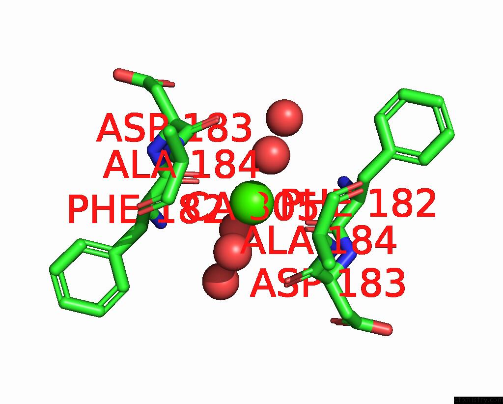
Mono view
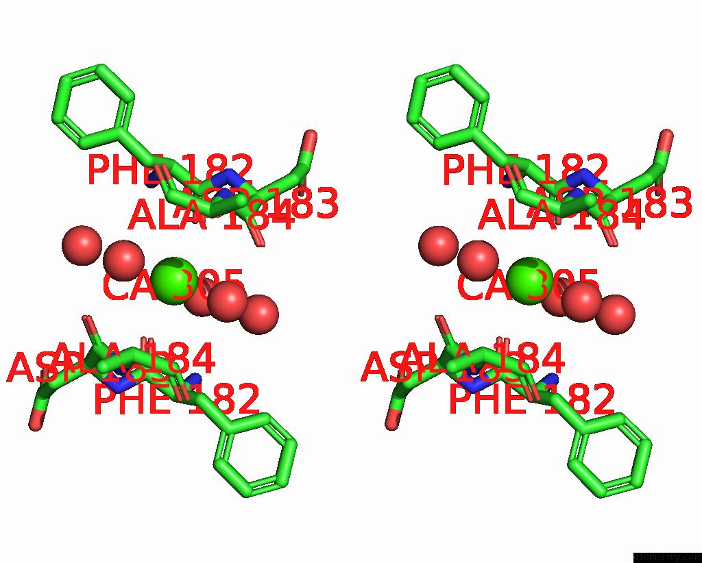
Stereo pair view
Mono view
Stereo pair view
A full contact list of Calcium with other atoms in the Ca binding
site number 1 of Crystal Structure of Cis-Epoxysuccinate Hydrolases Klcesh[L] Mutant D48N Complexed with L-Ta within 5.0Å range:
|
Calcium binding site 2 out of 2 in 8wbt
Go back to
Calcium binding site 2 out
of 2 in the Crystal Structure of Cis-Epoxysuccinate Hydrolases Klcesh[L] Mutant D48N Complexed with L-Ta
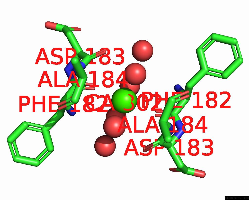
Mono view
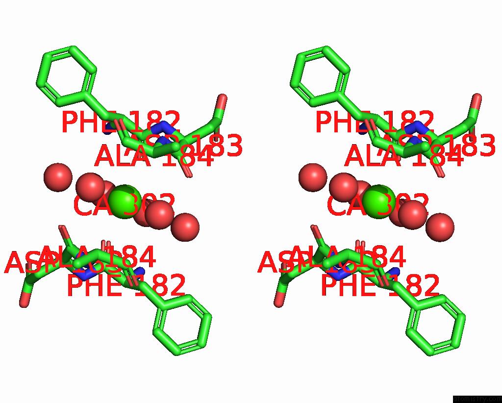
Stereo pair view
Mono view
Stereo pair view
A full contact list of Calcium with other atoms in the Ca binding
site number 2 of Crystal Structure of Cis-Epoxysuccinate Hydrolases Klcesh[L] Mutant D48N Complexed with L-Ta within 5.0Å range:
|
Reference:
S.Dong,
J.Xuan,
Y.Feng,
Q.Cui.
Deciphering the Stereo-Specific Catalytic Mechanisms of Cis-Epoxysuccinate Hydrolases Producing L(+)-Tartaric Acid. J.Biol.Chem. 05635 2024.
ISSN: ESSN 1083-351X
PubMed: 38199576
DOI: 10.1016/J.JBC.2024.105635
Page generated: Thu Jul 10 08:07:47 2025
ISSN: ESSN 1083-351X
PubMed: 38199576
DOI: 10.1016/J.JBC.2024.105635
Last articles
F in 7PX6F in 7PVU
F in 7PRV
F in 7PRW
F in 7PMQ
F in 7PRX
F in 7PPH
F in 7PRM
F in 7PQV
F in 7PQS