Calcium »
PDB 8x8g-8yib »
8x8g »
Calcium in PDB 8x8g: Crystal Structure of Endosz Mutant D234M, From Streptococcus Equi Subsp. Zooepidemicus SZ105, in Complex with Oligosaccharide G2S2- Oxazoline
Protein crystallography data
The structure of Crystal Structure of Endosz Mutant D234M, From Streptococcus Equi Subsp. Zooepidemicus SZ105, in Complex with Oligosaccharide G2S2- Oxazoline, PDB code: 8x8g
was solved by
H.H.Guan,
C.C.Lin,
Y.C.Hsieh,
C.J.Chen,
with X-Ray Crystallography technique. A brief refinement statistics is given in the table below:
| Resolution Low / High (Å) | 46.44 / 2.27 |
| Space group | P 21 21 21 |
| Cell size a, b, c (Å), α, β, γ (°) | 101.061, 232.603, 50.654, 90, 90, 90 |
| R / Rfree (%) | 20.8 / 24.9 |
Calcium Binding Sites:
The binding sites of Calcium atom in the Crystal Structure of Endosz Mutant D234M, From Streptococcus Equi Subsp. Zooepidemicus SZ105, in Complex with Oligosaccharide G2S2- Oxazoline
(pdb code 8x8g). This binding sites where shown within
5.0 Angstroms radius around Calcium atom.
In total only one binding site of Calcium was determined in the Crystal Structure of Endosz Mutant D234M, From Streptococcus Equi Subsp. Zooepidemicus SZ105, in Complex with Oligosaccharide G2S2- Oxazoline, PDB code: 8x8g:
In total only one binding site of Calcium was determined in the Crystal Structure of Endosz Mutant D234M, From Streptococcus Equi Subsp. Zooepidemicus SZ105, in Complex with Oligosaccharide G2S2- Oxazoline, PDB code: 8x8g:
Calcium binding site 1 out of 1 in 8x8g
Go back to
Calcium binding site 1 out
of 1 in the Crystal Structure of Endosz Mutant D234M, From Streptococcus Equi Subsp. Zooepidemicus SZ105, in Complex with Oligosaccharide G2S2- Oxazoline
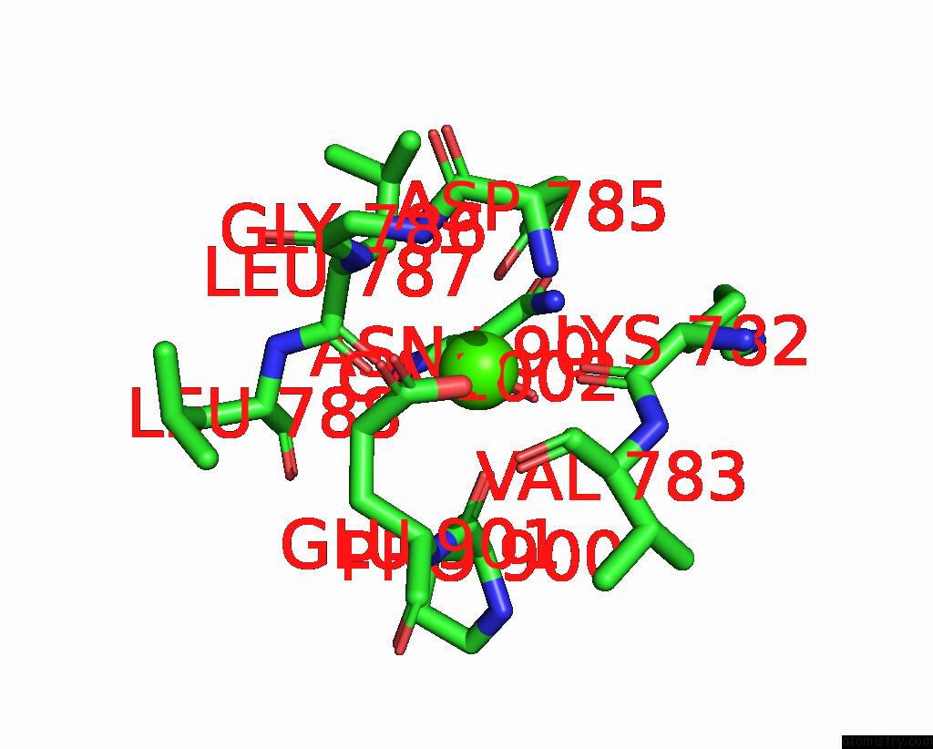
Mono view
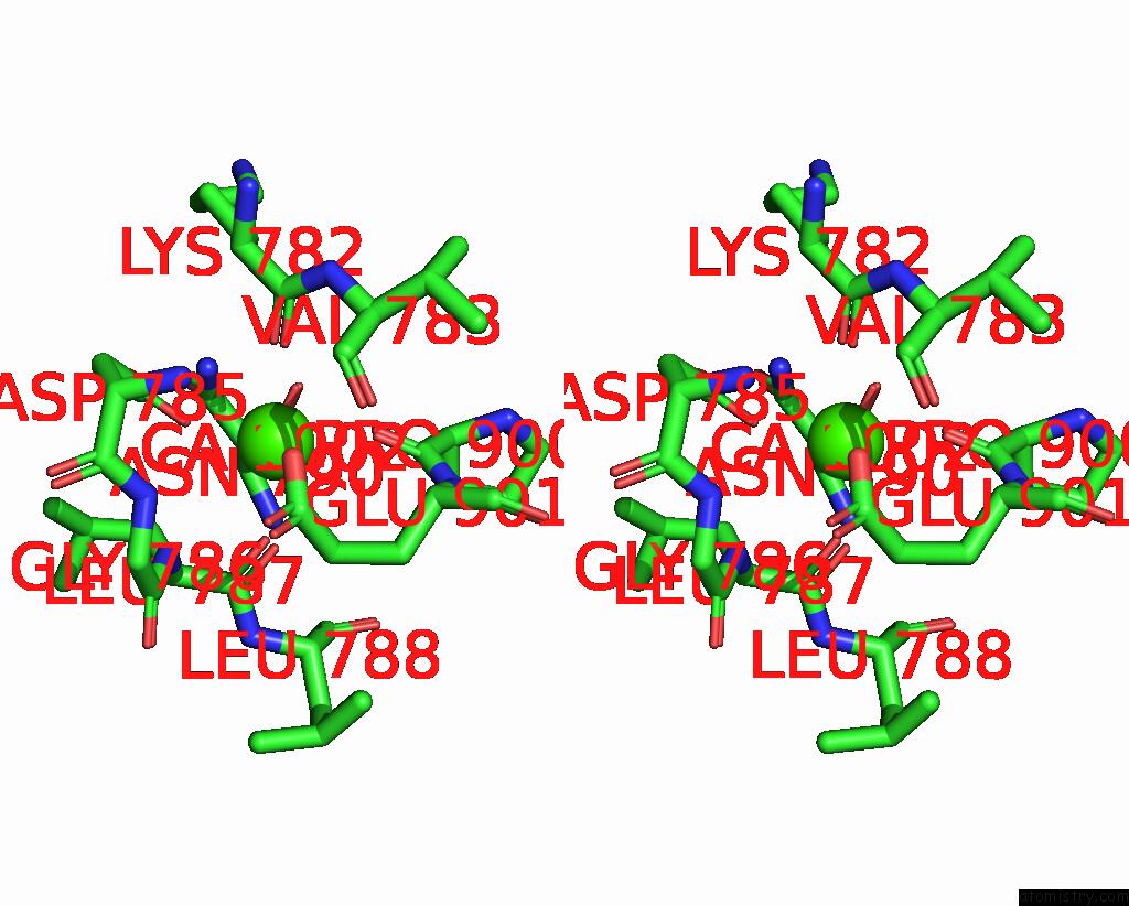
Stereo pair view
Mono view
Stereo pair view
A full contact list of Calcium with other atoms in the Ca binding
site number 1 of Crystal Structure of Endosz Mutant D234M, From Streptococcus Equi Subsp. Zooepidemicus SZ105, in Complex with Oligosaccharide G2S2- Oxazoline within 5.0Å range:
|
Reference:
Y.C.Hsieh,
H.H.Guan,
C.C.Lin,
T.Y.Huang,
P.Chuankhayan,
N.C.Chen,
N.H.Wang,
P.L.Hu,
Y.C.Tsai,
Y.C.Huang,
M.Yoshimura,
P.J.Lin,
Y.H.Hsieh,
C.J.Chen.
Structure-Based High-Efficiency Homogeneous Antibody Platform By Endoglycosidase Sz Provides Insights Into Its Transglycosylation Mechanism Jacs Au V. 4 2130 2024.
ISSN: ESSN 2691-3704
DOI: 10.1021/JACSAU.4C00004
Page generated: Thu Jul 10 08:19:43 2025
ISSN: ESSN 2691-3704
DOI: 10.1021/JACSAU.4C00004
Last articles
Fe in 2YXOFe in 2YRS
Fe in 2YXC
Fe in 2YNM
Fe in 2YVJ
Fe in 2YP1
Fe in 2YU2
Fe in 2YU1
Fe in 2YQB
Fe in 2YOO