Calcium »
PDB 8x8g-8yib »
8xrx »
Calcium in PDB 8xrx: The Crystal Structure of A GH3 Enzyme CCBGL3B with Glucose and Gentiobiose
Protein crystallography data
The structure of The Crystal Structure of A GH3 Enzyme CCBGL3B with Glucose and Gentiobiose, PDB code: 8xrx
was solved by
J.Y.Su,
with X-Ray Crystallography technique. A brief refinement statistics is given in the table below:
| Resolution Low / High (Å) | 19.95 / 2.50 |
| Space group | C 1 2 1 |
| Cell size a, b, c (Å), α, β, γ (°) | 163.095, 183.009, 269.154, 90, 100.59, 90 |
| R / Rfree (%) | 19.7 / 23 |
Calcium Binding Sites:
The binding sites of Calcium atom in the The Crystal Structure of A GH3 Enzyme CCBGL3B with Glucose and Gentiobiose
(pdb code 8xrx). This binding sites where shown within
5.0 Angstroms radius around Calcium atom.
In total 6 binding sites of Calcium where determined in the The Crystal Structure of A GH3 Enzyme CCBGL3B with Glucose and Gentiobiose, PDB code: 8xrx:
Jump to Calcium binding site number: 1; 2; 3; 4; 5; 6;
In total 6 binding sites of Calcium where determined in the The Crystal Structure of A GH3 Enzyme CCBGL3B with Glucose and Gentiobiose, PDB code: 8xrx:
Jump to Calcium binding site number: 1; 2; 3; 4; 5; 6;
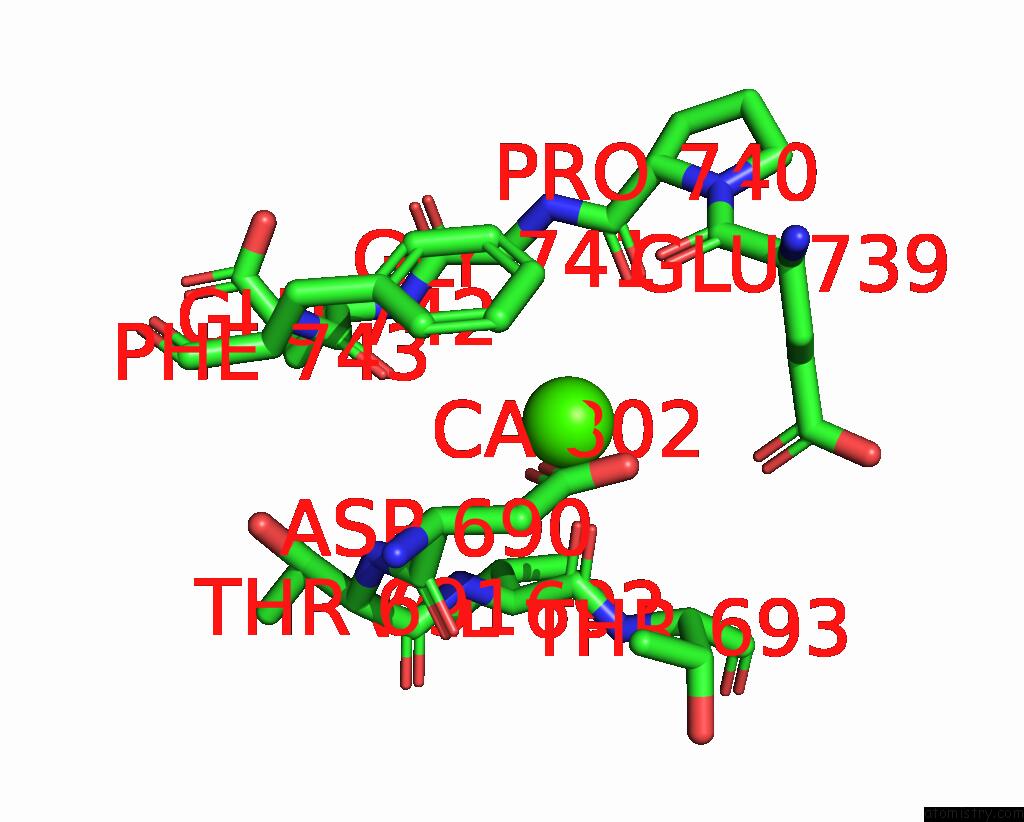
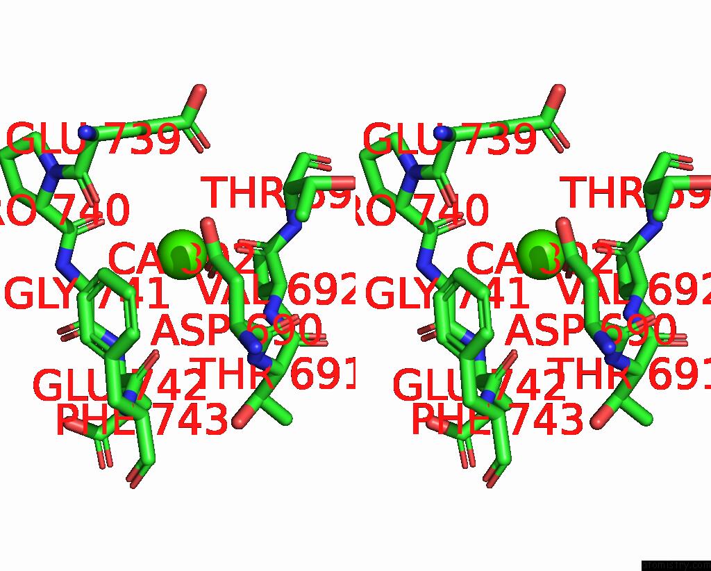
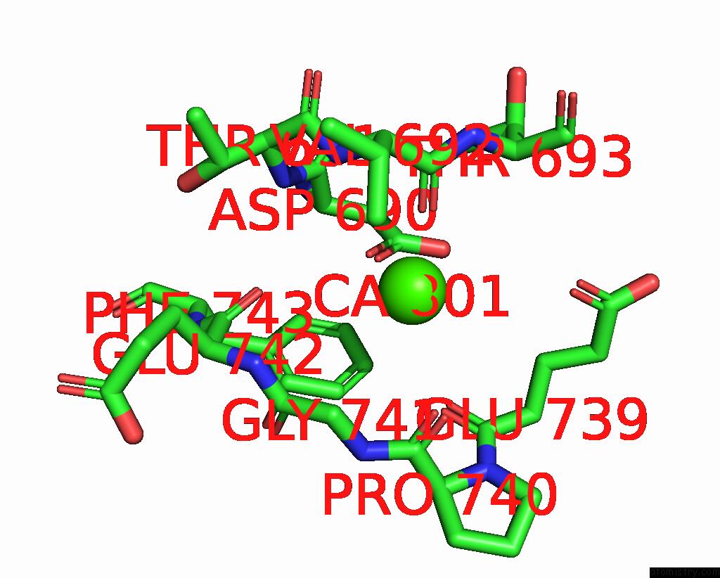
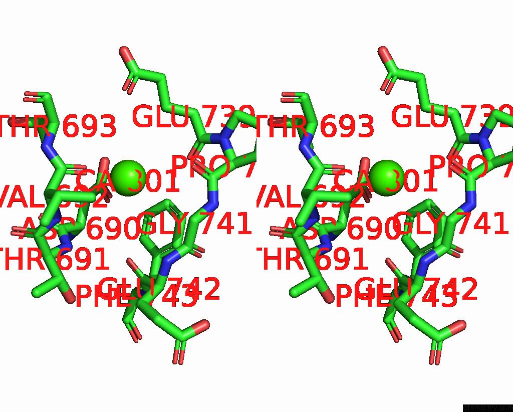
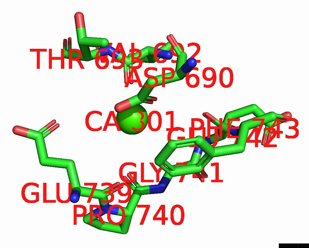
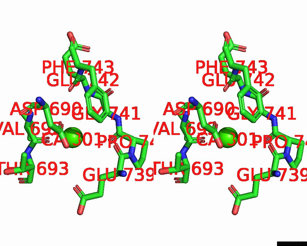
Calcium binding site 1 out of 6 in 8xrx
Go back to
Calcium binding site 1 out
of 6 in the The Crystal Structure of A GH3 Enzyme CCBGL3B with Glucose and Gentiobiose
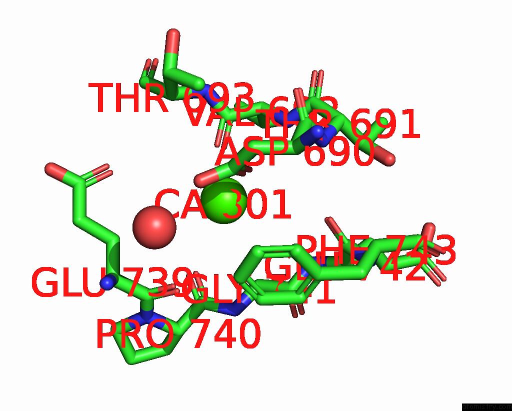
Mono view
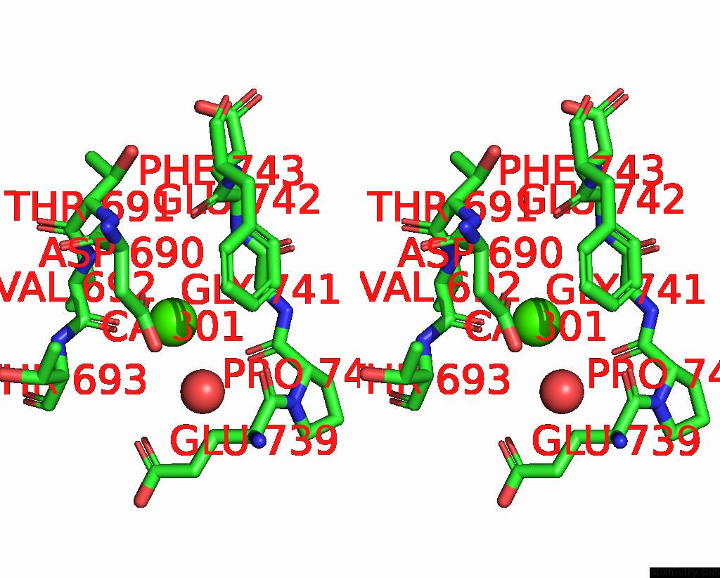
Stereo pair view
Mono view
Stereo pair view
A full contact list of Calcium with other atoms in the Ca binding
site number 1 of The Crystal Structure of A GH3 Enzyme CCBGL3B with Glucose and Gentiobiose within 5.0Å range:
|
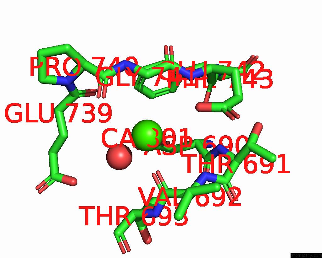
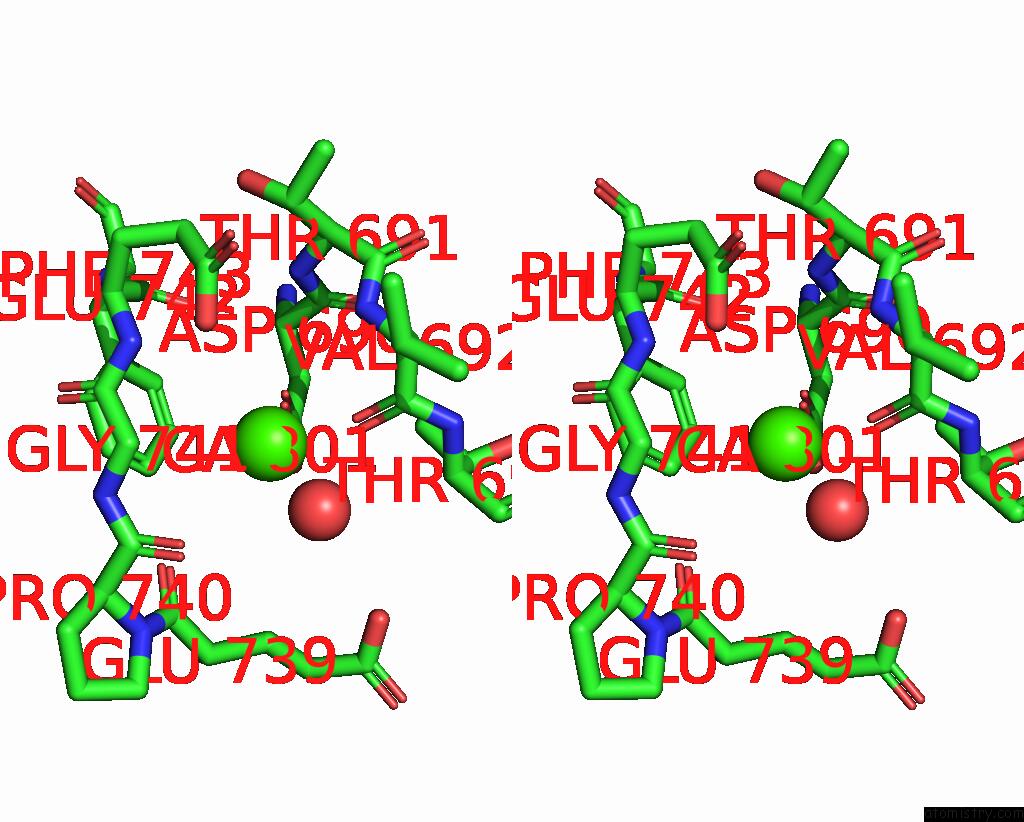
Calcium binding site 2 out of 6 in 8xrx
Go back to
Calcium binding site 2 out
of 6 in the The Crystal Structure of A GH3 Enzyme CCBGL3B with Glucose and Gentiobiose
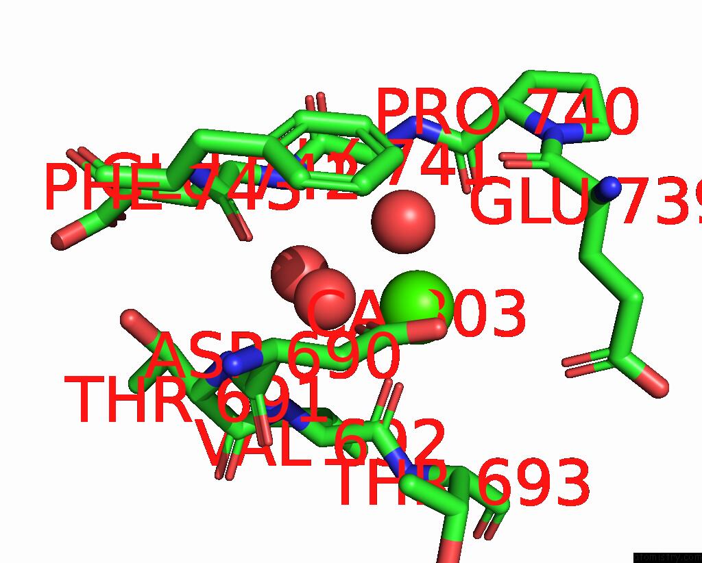
Mono view
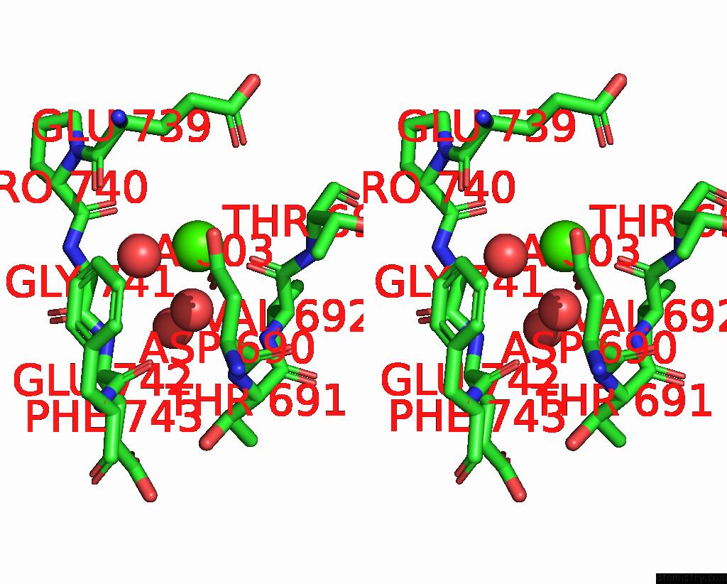
Stereo pair view
Mono view
Stereo pair view
A full contact list of Calcium with other atoms in the Ca binding
site number 2 of The Crystal Structure of A GH3 Enzyme CCBGL3B with Glucose and Gentiobiose within 5.0Å range:
|
Calcium binding site 3 out of 6 in 8xrx
Go back to
Calcium binding site 3 out
of 6 in the The Crystal Structure of A GH3 Enzyme CCBGL3B with Glucose and Gentiobiose
Mono view
Stereo pair view
Mono view
Stereo pair view
A full contact list of Calcium with other atoms in the Ca binding
site number 3 of The Crystal Structure of A GH3 Enzyme CCBGL3B with Glucose and Gentiobiose within 5.0Å range:
|
Calcium binding site 4 out of 6 in 8xrx
Go back to
Calcium binding site 4 out
of 6 in the The Crystal Structure of A GH3 Enzyme CCBGL3B with Glucose and Gentiobiose
Mono view
Stereo pair view
Mono view
Stereo pair view
A full contact list of Calcium with other atoms in the Ca binding
site number 4 of The Crystal Structure of A GH3 Enzyme CCBGL3B with Glucose and Gentiobiose within 5.0Å range:
|
Calcium binding site 5 out of 6 in 8xrx
Go back to
Calcium binding site 5 out
of 6 in the The Crystal Structure of A GH3 Enzyme CCBGL3B with Glucose and Gentiobiose
Mono view
Stereo pair view
Mono view
Stereo pair view
A full contact list of Calcium with other atoms in the Ca binding
site number 5 of The Crystal Structure of A GH3 Enzyme CCBGL3B with Glucose and Gentiobiose within 5.0Å range:
|
Calcium binding site 6 out of 6 in 8xrx
Go back to
Calcium binding site 6 out
of 6 in the The Crystal Structure of A GH3 Enzyme CCBGL3B with Glucose and Gentiobiose
Mono view
Stereo pair view
Mono view
Stereo pair view
A full contact list of Calcium with other atoms in the Ca binding
site number 6 of The Crystal Structure of A GH3 Enzyme CCBGL3B with Glucose and Gentiobiose within 5.0Å range:
|
Reference:
C.Hu,
Y.Wang,
W.Wang,
W.Cui,
X.Jia,
K.H.Mayo,
Y.Zhou,
J.Su,
Y.Yuan.
A Trapped Covalent Intermediate As A Key Catalytic Element in the Hydrolysis of A GH3 Beta-Glucosidase: An X-Ray Crystallographic and Biochemical Study. Int.J.Biol.Macromol. V. 265 31131 2024.
ISSN: ISSN 0141-8130
PubMed: 38527679
DOI: 10.1016/J.IJBIOMAC.2024.131131
Page generated: Thu Jul 10 08:27:56 2025
ISSN: ISSN 0141-8130
PubMed: 38527679
DOI: 10.1016/J.IJBIOMAC.2024.131131
Last articles
Fe in 2YXOFe in 2YRS
Fe in 2YXC
Fe in 2YNM
Fe in 2YVJ
Fe in 2YP1
Fe in 2YU2
Fe in 2YU1
Fe in 2YQB
Fe in 2YOO