Calcium »
PDB 8x8g-8yib »
8xx9 »
Calcium in PDB 8xx9: Rhodothermus Marinus Alpha-Amylase RMGH13_47A CBM48-A-B-C Domains
Enzymatic activity of Rhodothermus Marinus Alpha-Amylase RMGH13_47A CBM48-A-B-C Domains
All present enzymatic activity of Rhodothermus Marinus Alpha-Amylase RMGH13_47A CBM48-A-B-C Domains:
3.2.1.1;
3.2.1.1;
Protein crystallography data
The structure of Rhodothermus Marinus Alpha-Amylase RMGH13_47A CBM48-A-B-C Domains, PDB code: 8xx9
was solved by
T.Tonozuka,
with X-Ray Crystallography technique. A brief refinement statistics is given in the table below:
| Resolution Low / High (Å) | 43.50 / 1.55 |
| Space group | P 1 21 1 |
| Cell size a, b, c (Å), α, β, γ (°) | 43.77, 102.389, 63.397, 90, 96.42, 90 |
| R / Rfree (%) | 14.8 / 17.5 |
Other elements in 8xx9:
The structure of Rhodothermus Marinus Alpha-Amylase RMGH13_47A CBM48-A-B-C Domains also contains other interesting chemical elements:
| Manganese | (Mn) | 1 atom |
Calcium Binding Sites:
The binding sites of Calcium atom in the Rhodothermus Marinus Alpha-Amylase RMGH13_47A CBM48-A-B-C Domains
(pdb code 8xx9). This binding sites where shown within
5.0 Angstroms radius around Calcium atom.
In total only one binding site of Calcium was determined in the Rhodothermus Marinus Alpha-Amylase RMGH13_47A CBM48-A-B-C Domains, PDB code: 8xx9:
In total only one binding site of Calcium was determined in the Rhodothermus Marinus Alpha-Amylase RMGH13_47A CBM48-A-B-C Domains, PDB code: 8xx9:
Calcium binding site 1 out of 1 in 8xx9
Go back to
Calcium binding site 1 out
of 1 in the Rhodothermus Marinus Alpha-Amylase RMGH13_47A CBM48-A-B-C Domains
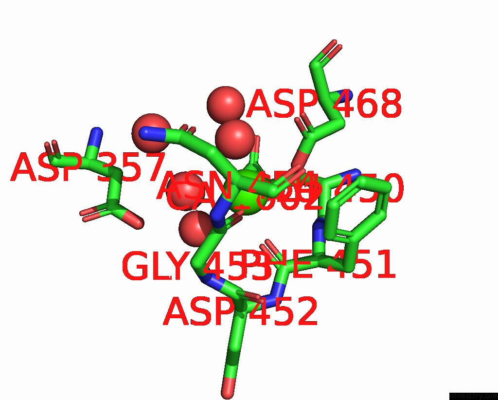
Mono view
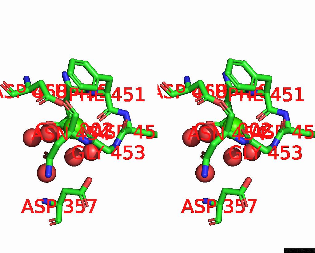
Stereo pair view
Mono view
Stereo pair view
A full contact list of Calcium with other atoms in the Ca binding
site number 1 of Rhodothermus Marinus Alpha-Amylase RMGH13_47A CBM48-A-B-C Domains within 5.0Å range:
|
Reference:
Y.Miyasaka,
K.Yokoyama,
T.Kozono,
Y.Kitano,
T.Miyazaki,
M.Sakaguchi,
A.Nishikawa,
T.Tonozuka.
Structural Basis For the Recognition of Alpha-1,6-Branched Alpha-Glucan By GH13_47 Alpha-Amylase From Rhodothermus Marinus To Be Published.
Page generated: Thu Jul 10 08:29:48 2025
Last articles
Cl in 8C5NCl in 8C5D
Cl in 8C41
Cl in 8C4N
Cl in 8C4O
Cl in 8C4D
Cl in 8C4M
Cl in 8C4J
Cl in 8BZL
Cl in 8C3L