Calcium »
PDB 8yj9-8zm6 »
8yk5 »
Calcium in PDB 8yk5: Structure of Glycerophosphoethanolamine Ethanolaminephosphodiesterase From Streptomyces Sanglieri
Enzymatic activity of Structure of Glycerophosphoethanolamine Ethanolaminephosphodiesterase From Streptomyces Sanglieri
All present enzymatic activity of Structure of Glycerophosphoethanolamine Ethanolaminephosphodiesterase From Streptomyces Sanglieri:
3.1.4.3;
3.1.4.3;
Protein crystallography data
The structure of Structure of Glycerophosphoethanolamine Ethanolaminephosphodiesterase From Streptomyces Sanglieri, PDB code: 8yk5
was solved by
K.Murayama,
D.Sugimori,
with X-Ray Crystallography technique. A brief refinement statistics is given in the table below:
| Resolution Low / High (Å) | 40.25 / 2.10 |
| Space group | C 1 2 1 |
| Cell size a, b, c (Å), α, β, γ (°) | 143.742, 179.184, 88.45, 90, 120.44, 90 |
| R / Rfree (%) | 18.3 / 21 |
Calcium Binding Sites:
The binding sites of Calcium atom in the Structure of Glycerophosphoethanolamine Ethanolaminephosphodiesterase From Streptomyces Sanglieri
(pdb code 8yk5). This binding sites where shown within
5.0 Angstroms radius around Calcium atom.
In total 2 binding sites of Calcium where determined in the Structure of Glycerophosphoethanolamine Ethanolaminephosphodiesterase From Streptomyces Sanglieri, PDB code: 8yk5:
Jump to Calcium binding site number: 1; 2;
In total 2 binding sites of Calcium where determined in the Structure of Glycerophosphoethanolamine Ethanolaminephosphodiesterase From Streptomyces Sanglieri, PDB code: 8yk5:
Jump to Calcium binding site number: 1; 2;
Calcium binding site 1 out of 2 in 8yk5
Go back to
Calcium binding site 1 out
of 2 in the Structure of Glycerophosphoethanolamine Ethanolaminephosphodiesterase From Streptomyces Sanglieri
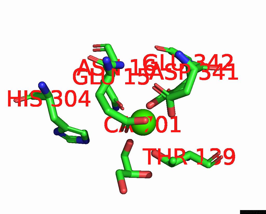
Mono view
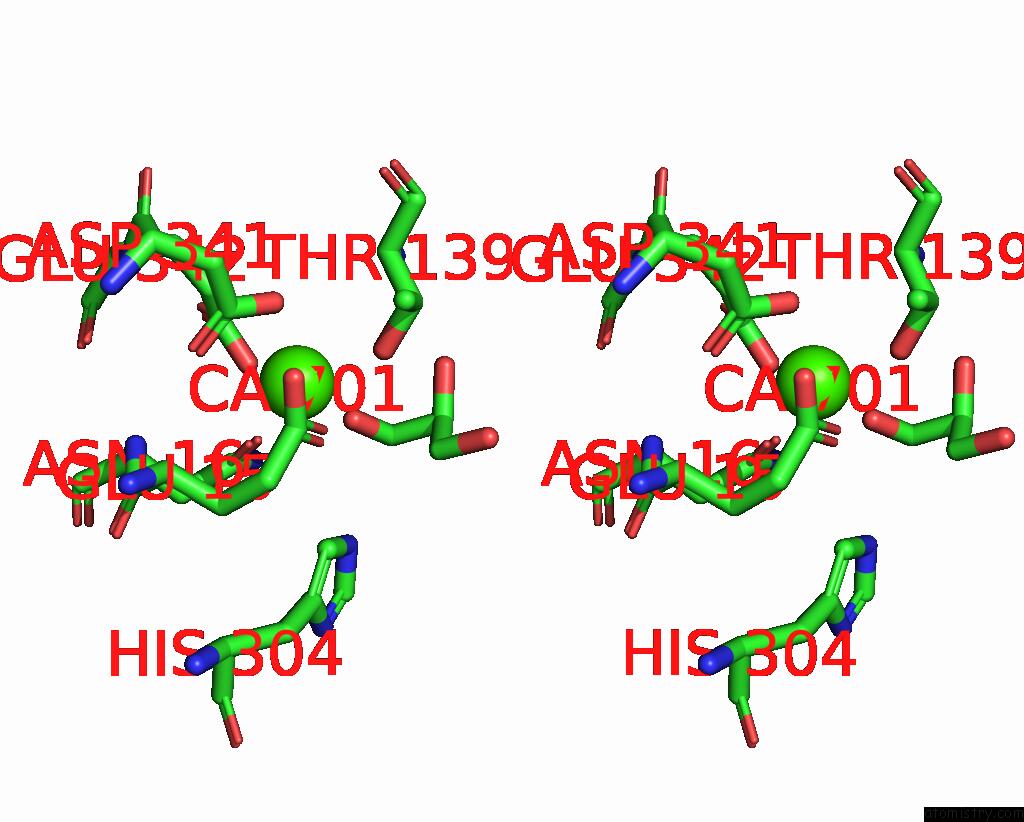
Stereo pair view
Mono view
Stereo pair view
A full contact list of Calcium with other atoms in the Ca binding
site number 1 of Structure of Glycerophosphoethanolamine Ethanolaminephosphodiesterase From Streptomyces Sanglieri within 5.0Å range:
|
Calcium binding site 2 out of 2 in 8yk5
Go back to
Calcium binding site 2 out
of 2 in the Structure of Glycerophosphoethanolamine Ethanolaminephosphodiesterase From Streptomyces Sanglieri
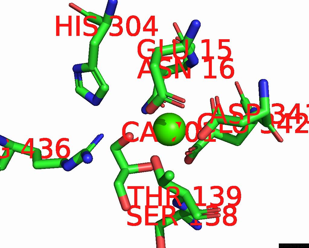
Mono view
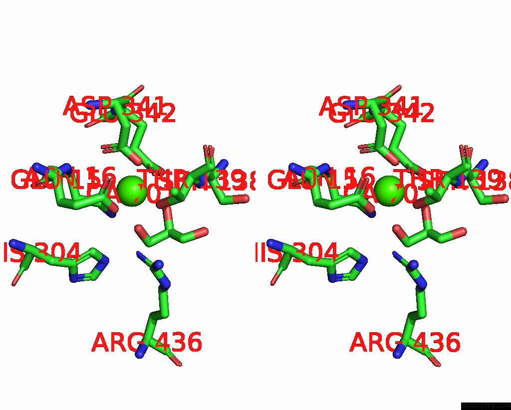
Stereo pair view
Mono view
Stereo pair view
A full contact list of Calcium with other atoms in the Ca binding
site number 2 of Structure of Glycerophosphoethanolamine Ethanolaminephosphodiesterase From Streptomyces Sanglieri within 5.0Å range:
|
Reference:
K.Murayama,
T.Hosaka,
M.Shirouzu,
D.Sugimori.
Structure of A Phosphodiesterase From Streptomyces Sanglieri with A Novel C-Terminal Domain. Biochem.Biophys.Res.Commun. V. 708 49784 2024.
ISSN: ESSN 1090-2104
PubMed: 38503170
DOI: 10.1016/J.BBRC.2024.149784
Page generated: Thu Jul 10 08:33:18 2025
ISSN: ESSN 1090-2104
PubMed: 38503170
DOI: 10.1016/J.BBRC.2024.149784
Last articles
Cl in 5L52Cl in 5L4H
Cl in 5L4E
Cl in 5L4I
Cl in 5L47
Cl in 5L3H
Cl in 5L3I
Cl in 5L2O
Cl in 5L3X
Cl in 5L39