Calcium »
PDB 8yj9-8zm6 »
8ywd »
Calcium in PDB 8ywd: Crystal Structure of Trehalose Synthase Mutant N253C From Deinococcus Radiodurans
Enzymatic activity of Crystal Structure of Trehalose Synthase Mutant N253C From Deinococcus Radiodurans
All present enzymatic activity of Crystal Structure of Trehalose Synthase Mutant N253C From Deinococcus Radiodurans:
5.4.99.16;
5.4.99.16;
Protein crystallography data
The structure of Crystal Structure of Trehalose Synthase Mutant N253C From Deinococcus Radiodurans, PDB code: 8ywd
was solved by
L.C.Ye,
S.C.Chen,
with X-Ray Crystallography technique. A brief refinement statistics is given in the table below:
| Resolution Low / High (Å) | 22.33 / 2.65 |
| Space group | P 1 21 1 |
| Cell size a, b, c (Å), α, β, γ (°) | 95.321, 196.852, 131.188, 90, 93.07, 90 |
| R / Rfree (%) | 17.2 / 23.4 |
Other elements in 8ywd:
The structure of Crystal Structure of Trehalose Synthase Mutant N253C From Deinococcus Radiodurans also contains other interesting chemical elements:
| Magnesium | (Mg) | 8 atoms |
Calcium Binding Sites:
The binding sites of Calcium atom in the Crystal Structure of Trehalose Synthase Mutant N253C From Deinococcus Radiodurans
(pdb code 8ywd). This binding sites where shown within
5.0 Angstroms radius around Calcium atom.
In total 8 binding sites of Calcium where determined in the Crystal Structure of Trehalose Synthase Mutant N253C From Deinococcus Radiodurans, PDB code: 8ywd:
Jump to Calcium binding site number: 1; 2; 3; 4; 5; 6; 7; 8;
In total 8 binding sites of Calcium where determined in the Crystal Structure of Trehalose Synthase Mutant N253C From Deinococcus Radiodurans, PDB code: 8ywd:
Jump to Calcium binding site number: 1; 2; 3; 4; 5; 6; 7; 8;
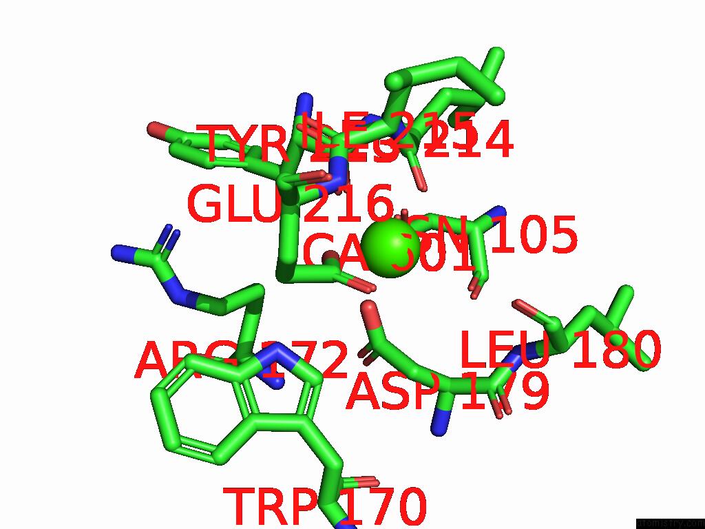
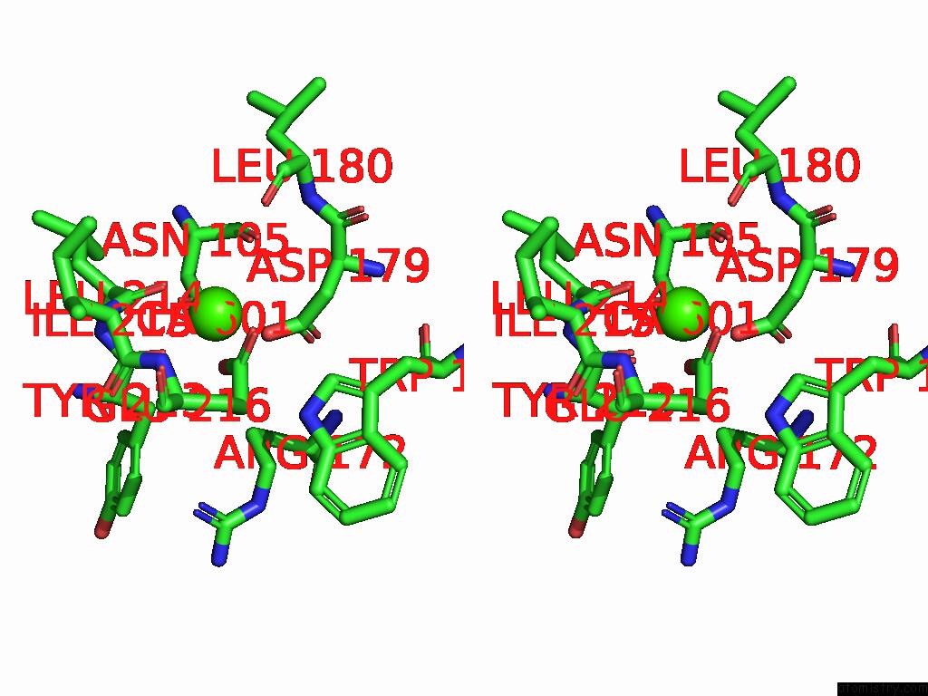
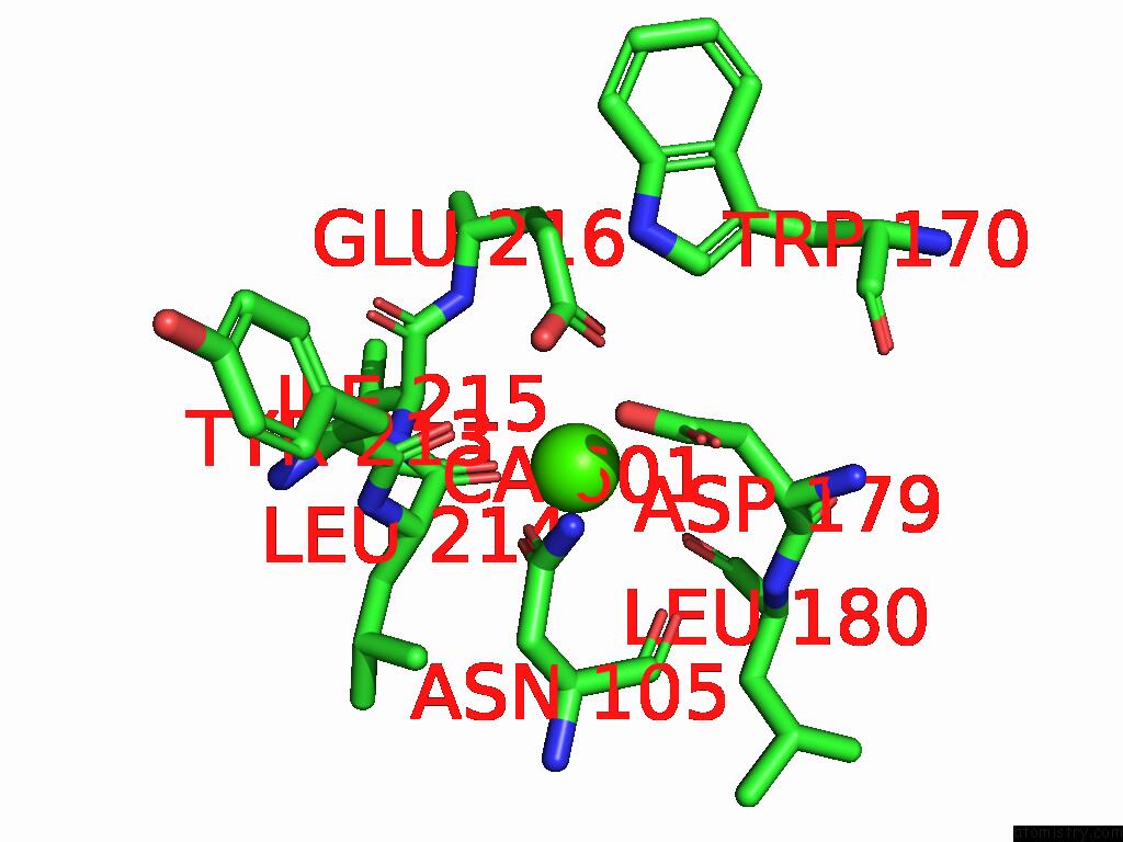
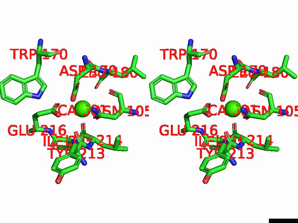
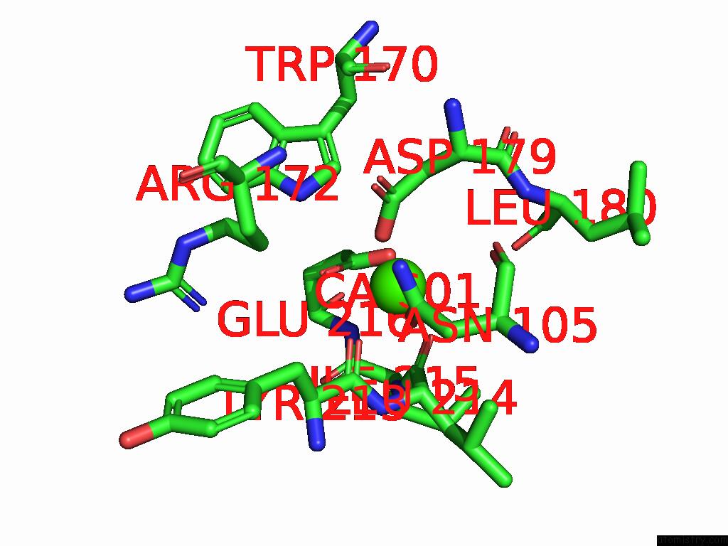
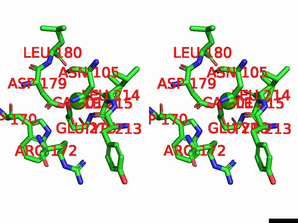
Calcium binding site 1 out of 8 in 8ywd
Go back to
Calcium binding site 1 out
of 8 in the Crystal Structure of Trehalose Synthase Mutant N253C From Deinococcus Radiodurans
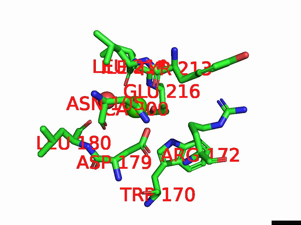
Mono view
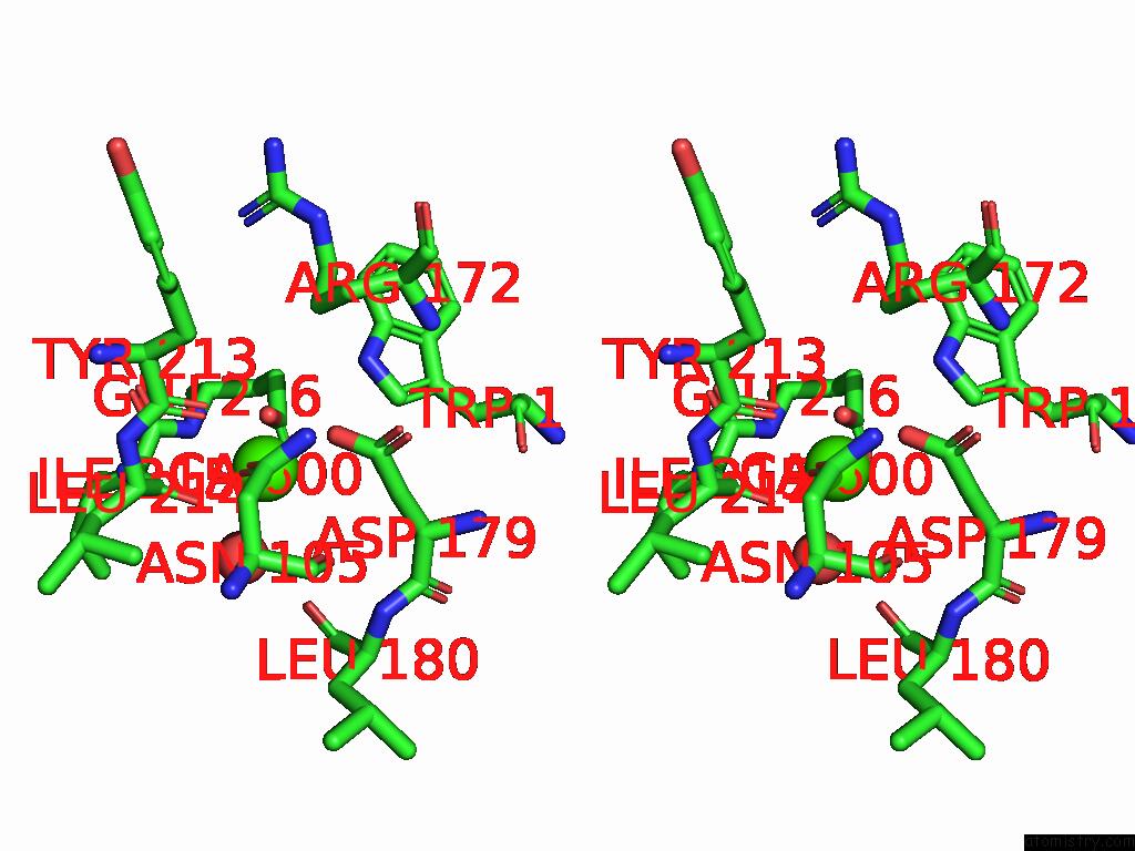
Stereo pair view
Mono view
Stereo pair view
A full contact list of Calcium with other atoms in the Ca binding
site number 1 of Crystal Structure of Trehalose Synthase Mutant N253C From Deinococcus Radiodurans within 5.0Å range:
|
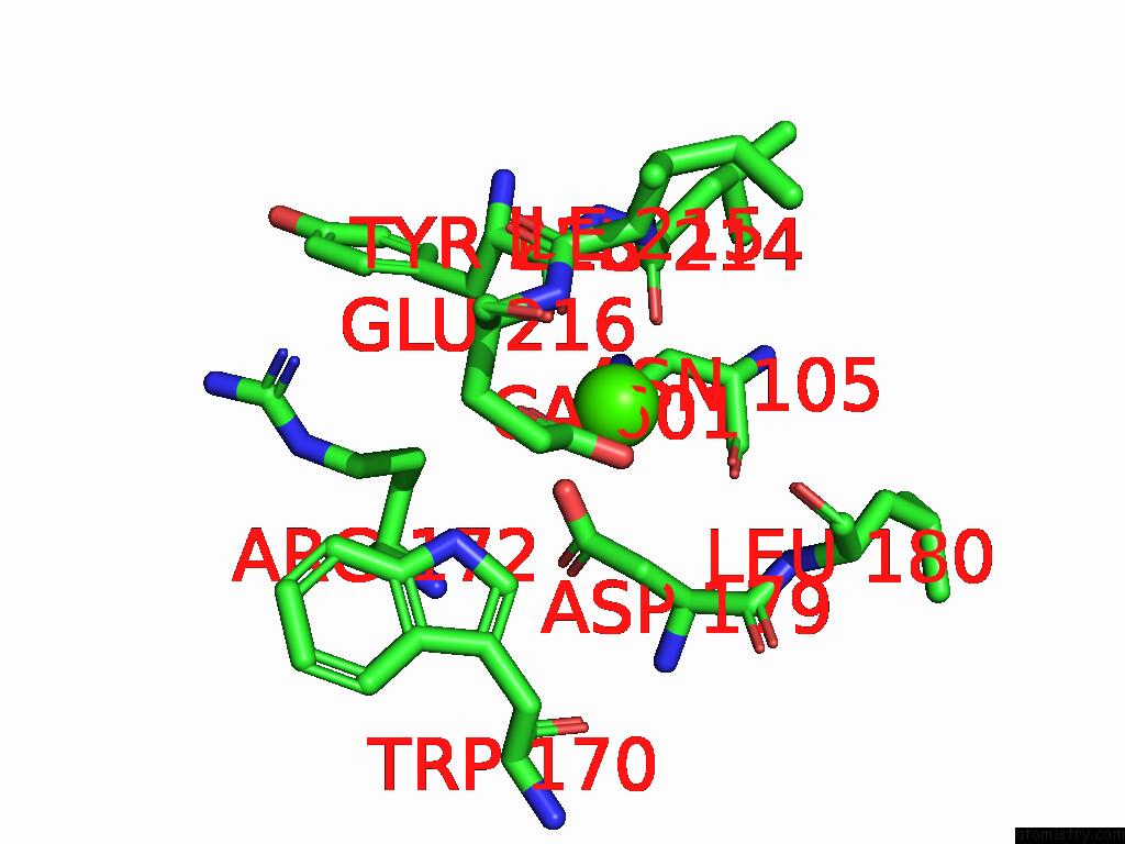
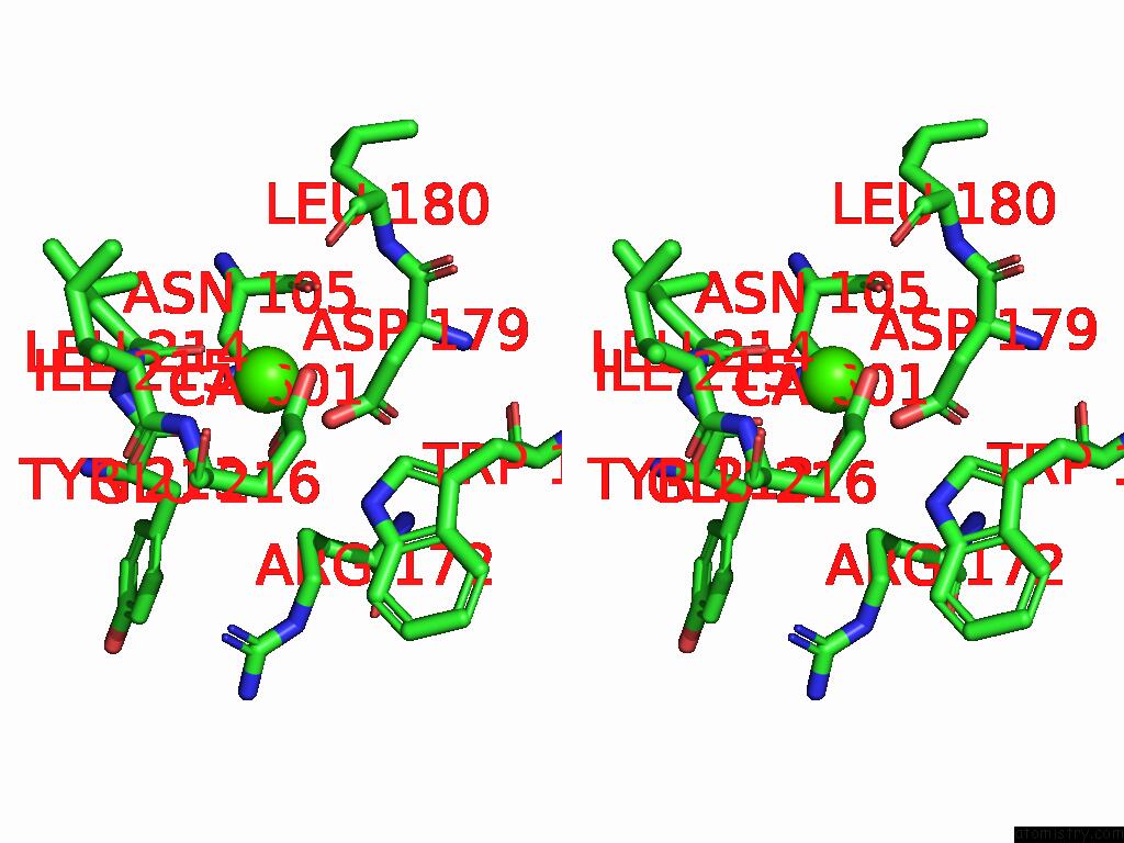
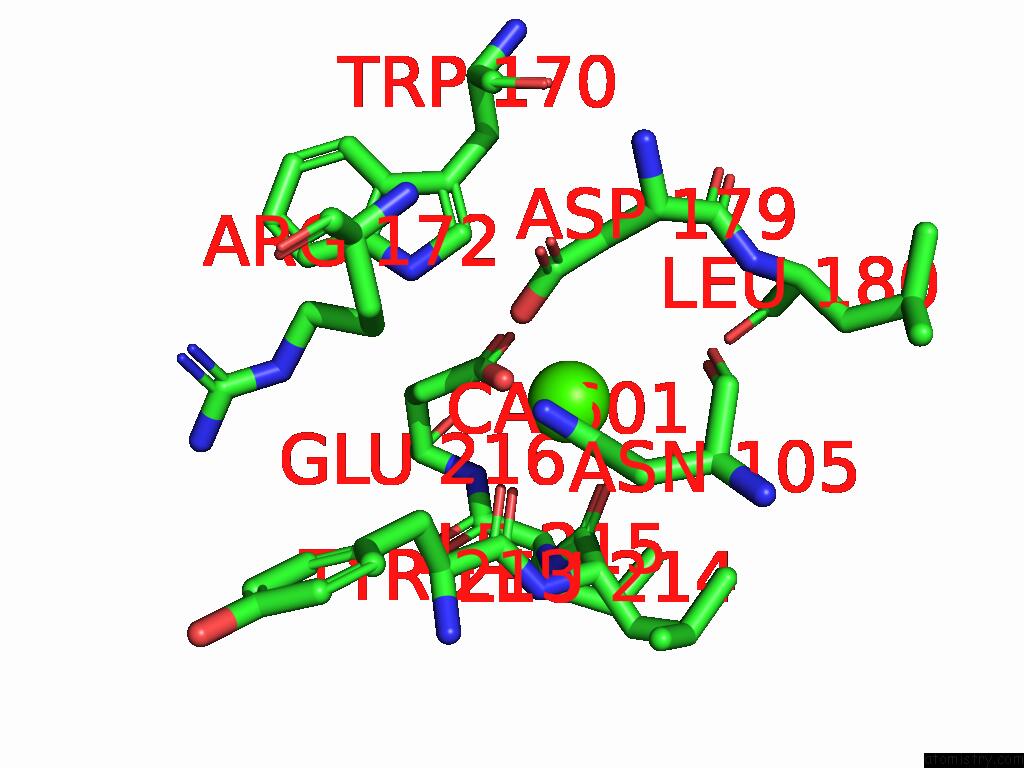
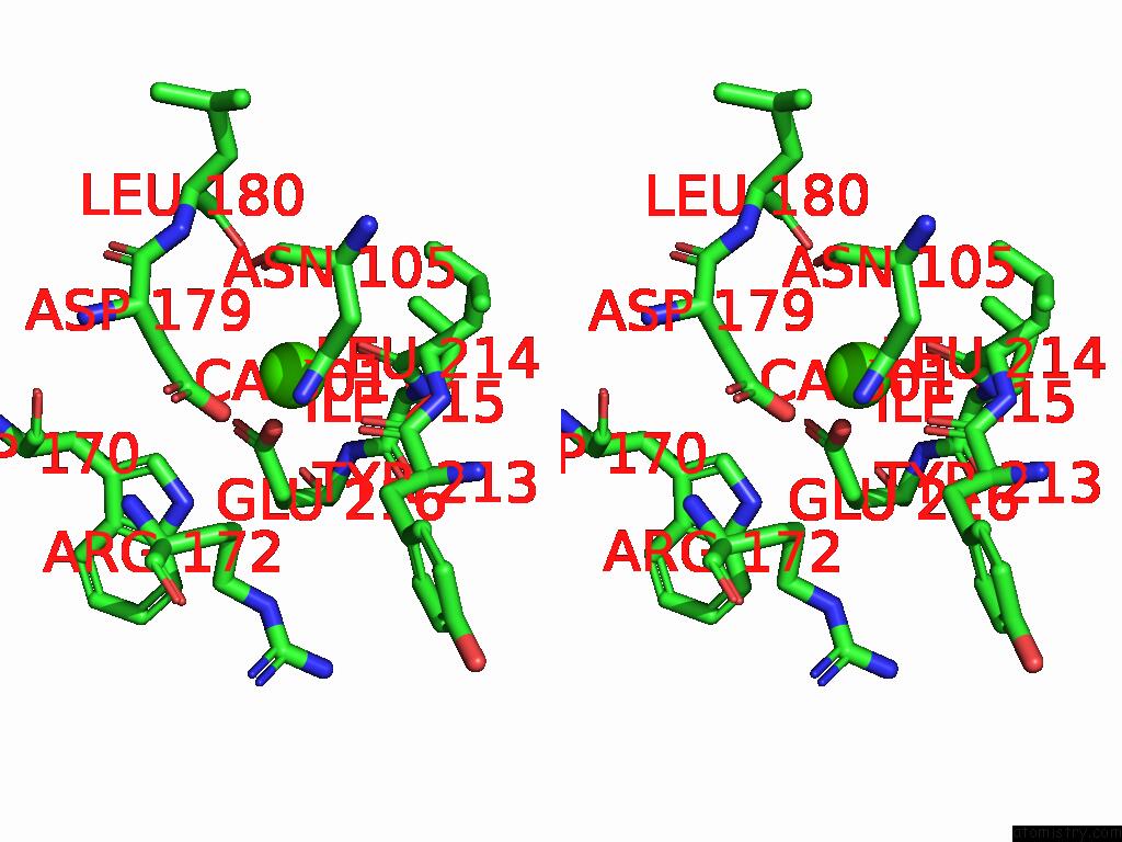
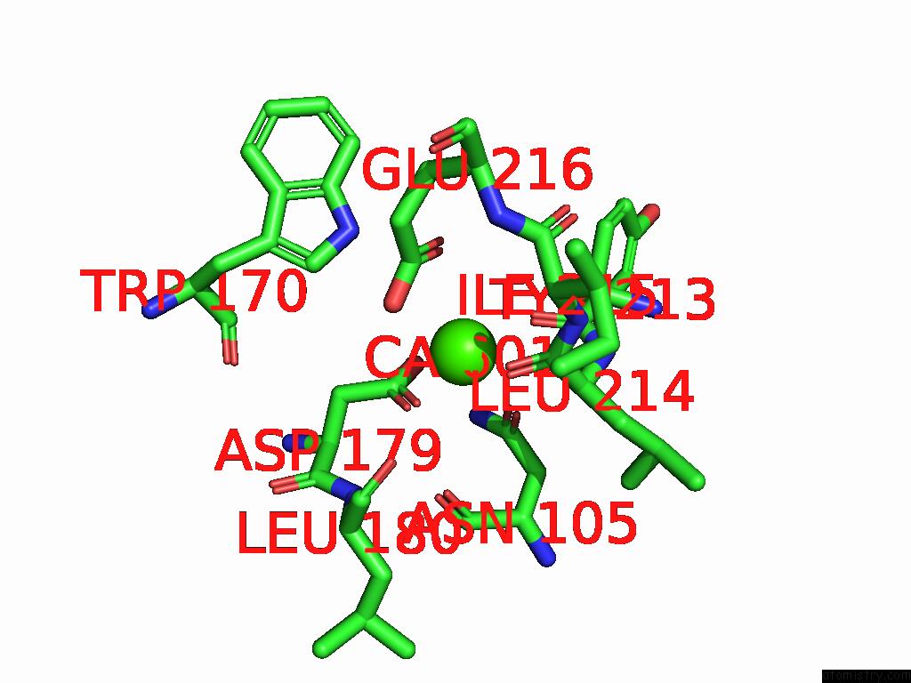
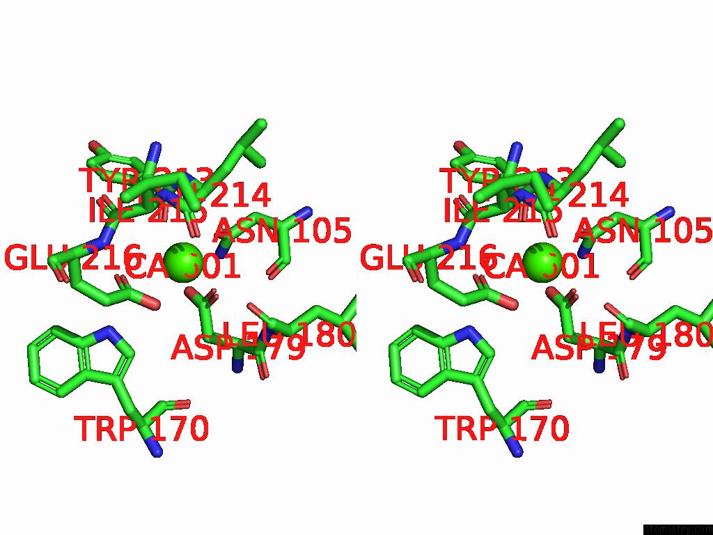
Calcium binding site 2 out of 8 in 8ywd
Go back to
Calcium binding site 2 out
of 8 in the Crystal Structure of Trehalose Synthase Mutant N253C From Deinococcus Radiodurans
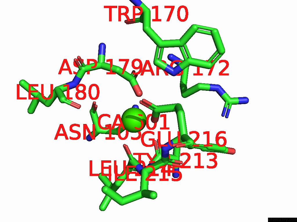
Mono view
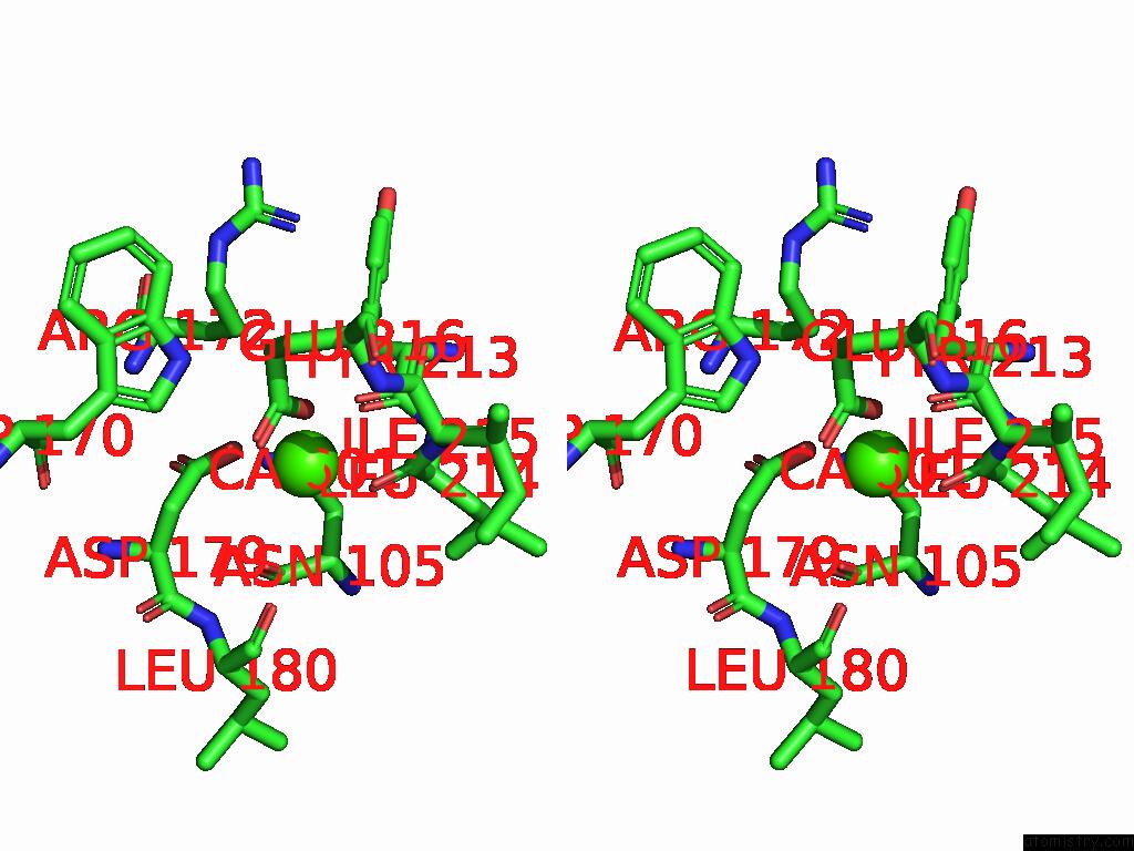
Stereo pair view
Mono view
Stereo pair view
A full contact list of Calcium with other atoms in the Ca binding
site number 2 of Crystal Structure of Trehalose Synthase Mutant N253C From Deinococcus Radiodurans within 5.0Å range:
|
Calcium binding site 3 out of 8 in 8ywd
Go back to
Calcium binding site 3 out
of 8 in the Crystal Structure of Trehalose Synthase Mutant N253C From Deinococcus Radiodurans
Mono view
Stereo pair view
Mono view
Stereo pair view
A full contact list of Calcium with other atoms in the Ca binding
site number 3 of Crystal Structure of Trehalose Synthase Mutant N253C From Deinococcus Radiodurans within 5.0Å range:
|
Calcium binding site 4 out of 8 in 8ywd
Go back to
Calcium binding site 4 out
of 8 in the Crystal Structure of Trehalose Synthase Mutant N253C From Deinococcus Radiodurans
Mono view
Stereo pair view
Mono view
Stereo pair view
A full contact list of Calcium with other atoms in the Ca binding
site number 4 of Crystal Structure of Trehalose Synthase Mutant N253C From Deinococcus Radiodurans within 5.0Å range:
|
Calcium binding site 5 out of 8 in 8ywd
Go back to
Calcium binding site 5 out
of 8 in the Crystal Structure of Trehalose Synthase Mutant N253C From Deinococcus Radiodurans
Mono view
Stereo pair view
Mono view
Stereo pair view
A full contact list of Calcium with other atoms in the Ca binding
site number 5 of Crystal Structure of Trehalose Synthase Mutant N253C From Deinococcus Radiodurans within 5.0Å range:
|
Calcium binding site 6 out of 8 in 8ywd
Go back to
Calcium binding site 6 out
of 8 in the Crystal Structure of Trehalose Synthase Mutant N253C From Deinococcus Radiodurans
Mono view
Stereo pair view
Mono view
Stereo pair view
A full contact list of Calcium with other atoms in the Ca binding
site number 6 of Crystal Structure of Trehalose Synthase Mutant N253C From Deinococcus Radiodurans within 5.0Å range:
|
Calcium binding site 7 out of 8 in 8ywd
Go back to
Calcium binding site 7 out
of 8 in the Crystal Structure of Trehalose Synthase Mutant N253C From Deinococcus Radiodurans
Mono view
Stereo pair view
Mono view
Stereo pair view
A full contact list of Calcium with other atoms in the Ca binding
site number 7 of Crystal Structure of Trehalose Synthase Mutant N253C From Deinococcus Radiodurans within 5.0Å range:
|
Calcium binding site 8 out of 8 in 8ywd
Go back to
Calcium binding site 8 out
of 8 in the Crystal Structure of Trehalose Synthase Mutant N253C From Deinococcus Radiodurans
Mono view
Stereo pair view
Mono view
Stereo pair view
A full contact list of Calcium with other atoms in the Ca binding
site number 8 of Crystal Structure of Trehalose Synthase Mutant N253C From Deinococcus Radiodurans within 5.0Å range:
|
Reference:
L.C.Ye,
S.Y.Chow,
S.C.Chang,
C.H.Kuo,
Y.L.Wang,
Y.J.Wei,
G.C.Lee,
S.H.Liaw,
W.M.Chen,
S.C.Chen.
Structural and Mutational Analyses of Trehalose Synthase From Deinococcus Radiodurans Reveal the Interconversion of Maltose-Trehalose Mechanism. J.Agric.Food Chem. V. 72 18649 2024.
ISSN: ESSN 1520-5118
PubMed: 39109746
DOI: 10.1021/ACS.JAFC.4C03661
Page generated: Thu Jul 10 08:37:25 2025
ISSN: ESSN 1520-5118
PubMed: 39109746
DOI: 10.1021/ACS.JAFC.4C03661
Last articles
Cl in 5L3XCl in 5L39
Cl in 5L38
Cl in 5L35
Cl in 5L2U
Cl in 5L2N
Cl in 5L1E
Cl in 5L2K
Cl in 5L0K
Cl in 5L2M