Calcium »
PDB 8yj9-8zm6 »
8z3f »
Calcium in PDB 8z3f: Complex Structure of CIB2 and TMC1 CR1
Protein crystallography data
The structure of Complex Structure of CIB2 and TMC1 CR1, PDB code: 8z3f
was solved by
S.Wu,
L.Lin,
Q.Lu,
with X-Ray Crystallography technique. A brief refinement statistics is given in the table below:
| Resolution Low / High (Å) | 25.52 / 1.74 |
| Space group | C 1 2 1 |
| Cell size a, b, c (Å), α, β, γ (°) | 109.177, 36.225, 63.496, 90, 98.87, 90 |
| R / Rfree (%) | 17.7 / 20 |
Calcium Binding Sites:
The binding sites of Calcium atom in the Complex Structure of CIB2 and TMC1 CR1
(pdb code 8z3f). This binding sites where shown within
5.0 Angstroms radius around Calcium atom.
In total 3 binding sites of Calcium where determined in the Complex Structure of CIB2 and TMC1 CR1, PDB code: 8z3f:
Jump to Calcium binding site number: 1; 2; 3;
In total 3 binding sites of Calcium where determined in the Complex Structure of CIB2 and TMC1 CR1, PDB code: 8z3f:
Jump to Calcium binding site number: 1; 2; 3;
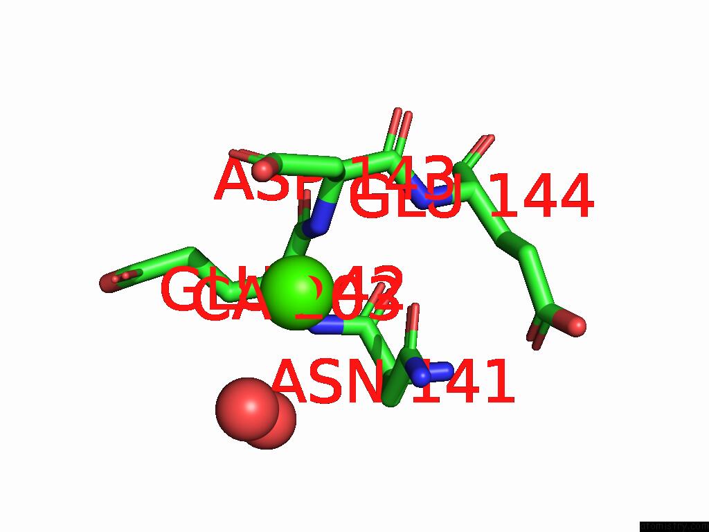
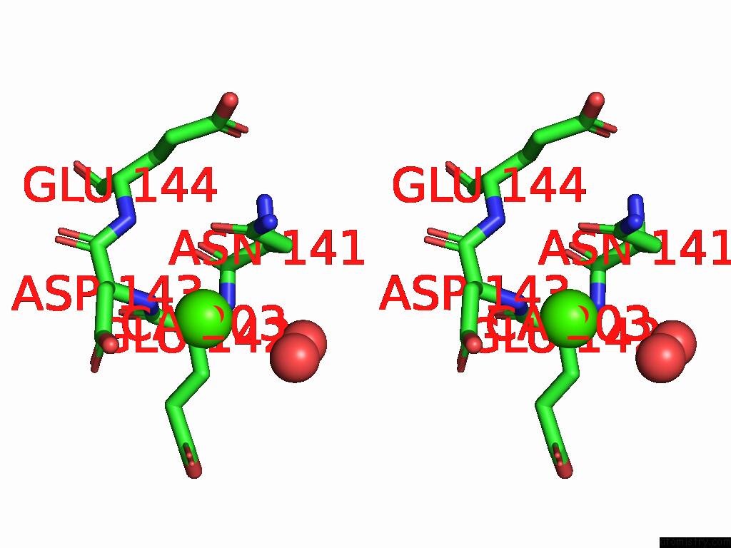
Calcium binding site 1 out of 3 in 8z3f
Go back to
Calcium binding site 1 out
of 3 in the Complex Structure of CIB2 and TMC1 CR1
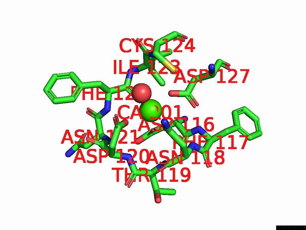
Mono view
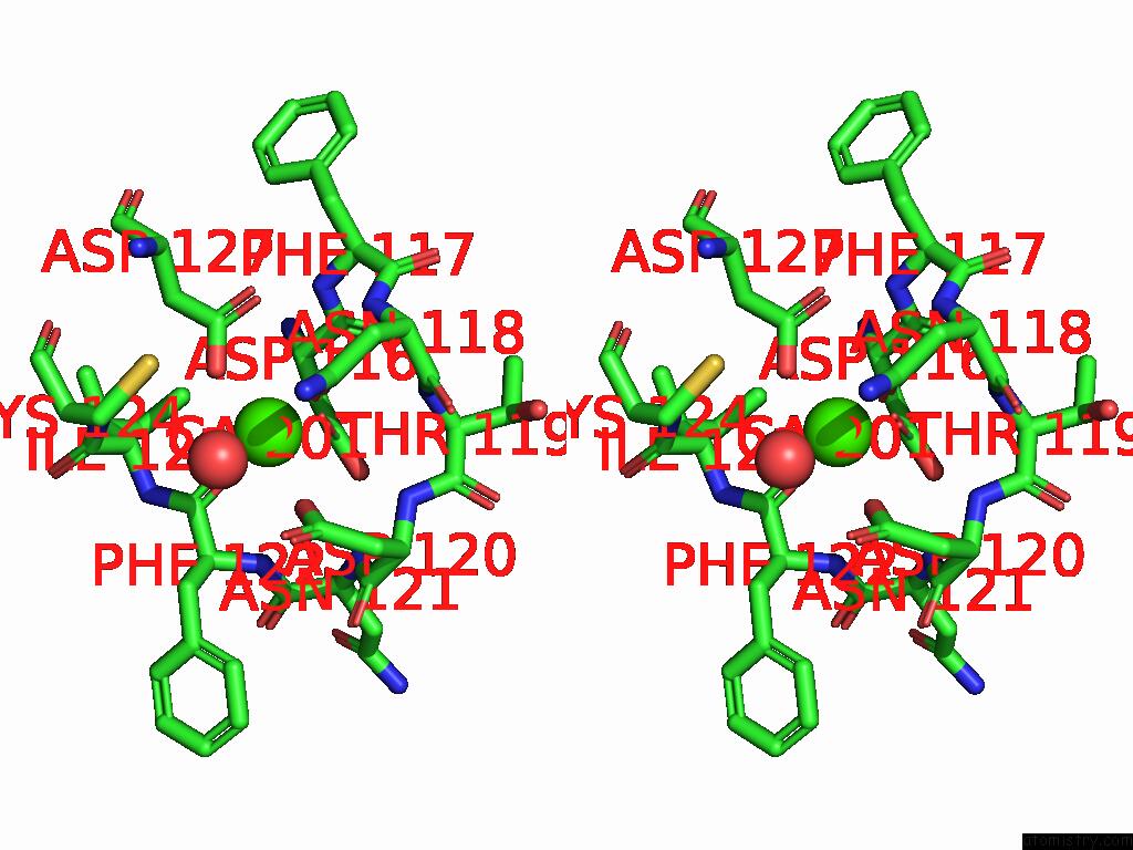
Stereo pair view
Mono view
Stereo pair view
A full contact list of Calcium with other atoms in the Ca binding
site number 1 of Complex Structure of CIB2 and TMC1 CR1 within 5.0Å range:
|
Calcium binding site 2 out of 3 in 8z3f
Go back to
Calcium binding site 2 out
of 3 in the Complex Structure of CIB2 and TMC1 CR1
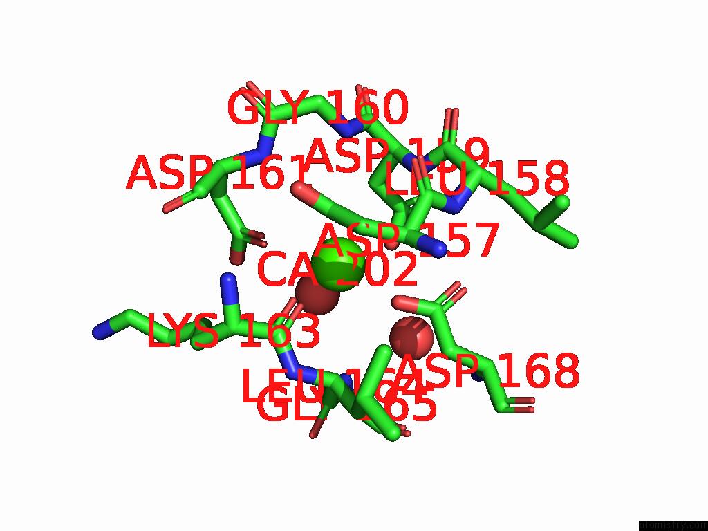
Mono view
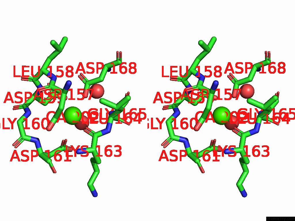
Stereo pair view
Mono view
Stereo pair view
A full contact list of Calcium with other atoms in the Ca binding
site number 2 of Complex Structure of CIB2 and TMC1 CR1 within 5.0Å range:
|
Calcium binding site 3 out of 3 in 8z3f
Go back to
Calcium binding site 3 out
of 3 in the Complex Structure of CIB2 and TMC1 CR1
Mono view
Stereo pair view
Mono view
Stereo pair view
A full contact list of Calcium with other atoms in the Ca binding
site number 3 of Complex Structure of CIB2 and TMC1 CR1 within 5.0Å range:
|
Reference:
S.Wu,
L.Lin,
Q.Hu,
X.Yao,
H.Wang,
S.Liu,
Q.Liu,
Y.Xi,
Y.Lin,
J.Gong,
R.Hu,
W.Zhan,
Y.Luo,
G.He,
Z.Liu,
W.Xiong,
Q.Wang,
Z.Xu,
F.Bai,
Q.Lu.
Mechano-Electrical Transduction Components TMC1-CIB2 Undergo A Ca 2+ -Induced Conformational Change Linked to Hearing Loss. Dev.Cell 2025.
ISSN: ISSN 1534-5807
PubMed: 39889697
DOI: 10.1016/J.DEVCEL.2025.01.004
Page generated: Thu Jul 10 08:42:15 2025
ISSN: ISSN 1534-5807
PubMed: 39889697
DOI: 10.1016/J.DEVCEL.2025.01.004
Last articles
Fe in 2YXOFe in 2YRS
Fe in 2YXC
Fe in 2YNM
Fe in 2YVJ
Fe in 2YP1
Fe in 2YU2
Fe in 2YU1
Fe in 2YQB
Fe in 2YOO