Calcium »
PDB 8znz-9awu »
8zrz »
Calcium in PDB 8zrz: The 1.26 Angstrom Resolution Structure of Bacillus Cereus Beta-Amylase in Complex with Maltose
Enzymatic activity of The 1.26 Angstrom Resolution Structure of Bacillus Cereus Beta-Amylase in Complex with Maltose
All present enzymatic activity of The 1.26 Angstrom Resolution Structure of Bacillus Cereus Beta-Amylase in Complex with Maltose:
3.2.1.2;
3.2.1.2;
Protein crystallography data
The structure of The 1.26 Angstrom Resolution Structure of Bacillus Cereus Beta-Amylase in Complex with Maltose, PDB code: 8zrz
was solved by
B.Mikami,
A.Hirata,
with X-Ray Crystallography technique. A brief refinement statistics is given in the table below:
| Resolution Low / High (Å) | 10.00 / 1.26 |
| Space group | P 1 21 1 |
| Cell size a, b, c (Å), α, β, γ (°) | 56.974, 89.843, 66.035, 90, 103.09, 90 |
| R / Rfree (%) | n/a / n/a |
Calcium Binding Sites:
The binding sites of Calcium atom in the The 1.26 Angstrom Resolution Structure of Bacillus Cereus Beta-Amylase in Complex with Maltose
(pdb code 8zrz). This binding sites where shown within
5.0 Angstroms radius around Calcium atom.
In total only one binding site of Calcium was determined in the The 1.26 Angstrom Resolution Structure of Bacillus Cereus Beta-Amylase in Complex with Maltose, PDB code: 8zrz:
In total only one binding site of Calcium was determined in the The 1.26 Angstrom Resolution Structure of Bacillus Cereus Beta-Amylase in Complex with Maltose, PDB code: 8zrz:
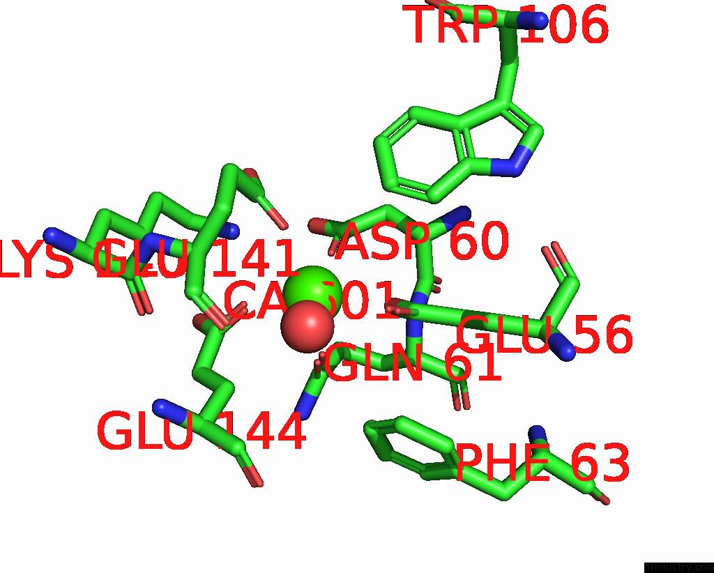
Calcium binding site 1 out of 1 in 8zrz
Go back to
Calcium binding site 1 out
of 1 in the The 1.26 Angstrom Resolution Structure of Bacillus Cereus Beta-Amylase in Complex with Maltose
Mono view

Stereo pair view
Mono view
Stereo pair view
A full contact list of Calcium with other atoms in the Ca binding
site number 1 of The 1.26 Angstrom Resolution Structure of Bacillus Cereus Beta-Amylase in Complex with Maltose within 5.0Å range:
|
Reference:
A.Hirata,
B.Mikami.
Structural Insight Into Sugar-Binding Modes of Microbial Beta-Amylase. Biochem.Biophys.Res.Commun. V. 733 2024.
ISSN: ESSN 1090-2104
DOI: 10.1016/J.BBRC.2024.150695
Page generated: Thu Jul 10 08:47:52 2025
ISSN: ESSN 1090-2104
DOI: 10.1016/J.BBRC.2024.150695
Last articles
Cl in 5QBZCl in 5QBW
Cl in 5QB4
Cl in 5QB3
Cl in 5QB1
Cl in 5QB2
Cl in 5QB0
Cl in 5QAZ
Cl in 5QAY
Cl in 5QAX