Calcium »
PDB 8znz-9awu »
9awo »
Calcium in PDB 9awo: Crystal Structure of Trypsin at 250 Kelvin with Benzamidine (Duplicate)
Enzymatic activity of Crystal Structure of Trypsin at 250 Kelvin with Benzamidine (Duplicate)
All present enzymatic activity of Crystal Structure of Trypsin at 250 Kelvin with Benzamidine (Duplicate):
3.4.21.4;
3.4.21.4;
Protein crystallography data
The structure of Crystal Structure of Trypsin at 250 Kelvin with Benzamidine (Duplicate), PDB code: 9awo
was solved by
L.M.T.R.Lima,
F.De Sa Ribeiro,
with X-Ray Crystallography technique. A brief refinement statistics is given in the table below:
| Resolution Low / High (Å) | 26.93 / 1.50 |
| Space group | P 21 21 21 |
| Cell size a, b, c (Å), α, β, γ (°) | 53.824, 56.986, 66.449, 90, 90, 90 |
| R / Rfree (%) | 18.2 / 20.6 |
Calcium Binding Sites:
The binding sites of Calcium atom in the Crystal Structure of Trypsin at 250 Kelvin with Benzamidine (Duplicate)
(pdb code 9awo). This binding sites where shown within
5.0 Angstroms radius around Calcium atom.
In total only one binding site of Calcium was determined in the Crystal Structure of Trypsin at 250 Kelvin with Benzamidine (Duplicate), PDB code: 9awo:
In total only one binding site of Calcium was determined in the Crystal Structure of Trypsin at 250 Kelvin with Benzamidine (Duplicate), PDB code: 9awo:
Calcium binding site 1 out of 1 in 9awo
Go back to
Calcium binding site 1 out
of 1 in the Crystal Structure of Trypsin at 250 Kelvin with Benzamidine (Duplicate)
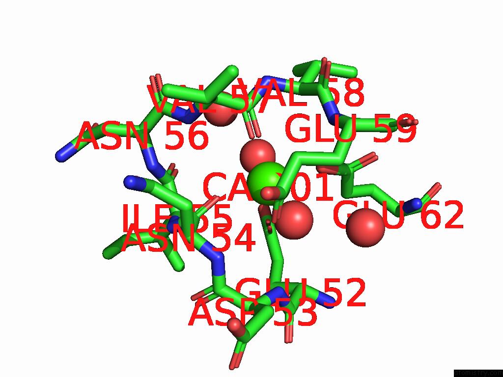
Mono view
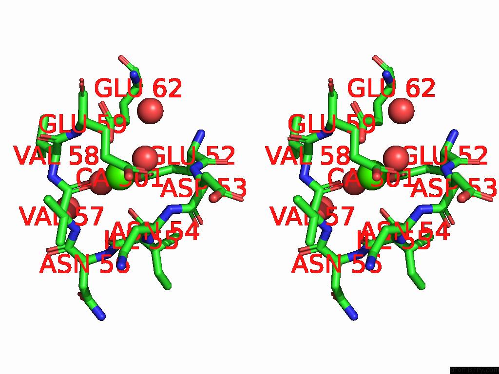
Stereo pair view
Mono view
Stereo pair view
A full contact list of Calcium with other atoms in the Ca binding
site number 1 of Crystal Structure of Trypsin at 250 Kelvin with Benzamidine (Duplicate) within 5.0Å range:
|
Reference:
L.M.T.R.Lima,
F.De Sa Ribeiro.
Crystal Structure of Trypsin at 250 Kelvin with Benzamidine (Duplicate) To Be Published.
Page generated: Thu Jul 10 08:51:02 2025
Last articles
Cl in 5J8ZCl in 5J8F
Cl in 5J8I
Cl in 5J8C
Cl in 5J7S
Cl in 5J7R
Cl in 5J7F
Cl in 5J72
Cl in 5J6B
Cl in 5J5Y