Calcium »
PDB 9bi0-9ctr »
9buj »
Calcium in PDB 9buj: Structure of PFPL1 From Pseudoalteromonas Fuliginea
Protein crystallography data
The structure of Structure of PFPL1 From Pseudoalteromonas Fuliginea, PDB code: 9buj
was solved by
A.B.Boraston,
J.K.Hobbs,
with X-Ray Crystallography technique. A brief refinement statistics is given in the table below:
| Resolution Low / High (Å) | 19.50 / 2.19 |
| Space group | P 21 21 21 |
| Cell size a, b, c (Å), α, β, γ (°) | 48.504, 58.137, 149.08, 90, 90, 90 |
| R / Rfree (%) | 18.2 / 25.2 |
Calcium Binding Sites:
The binding sites of Calcium atom in the Structure of PFPL1 From Pseudoalteromonas Fuliginea
(pdb code 9buj). This binding sites where shown within
5.0 Angstroms radius around Calcium atom.
In total only one binding site of Calcium was determined in the Structure of PFPL1 From Pseudoalteromonas Fuliginea, PDB code: 9buj:
In total only one binding site of Calcium was determined in the Structure of PFPL1 From Pseudoalteromonas Fuliginea, PDB code: 9buj:
Calcium binding site 1 out of 1 in 9buj
Go back to
Calcium binding site 1 out
of 1 in the Structure of PFPL1 From Pseudoalteromonas Fuliginea
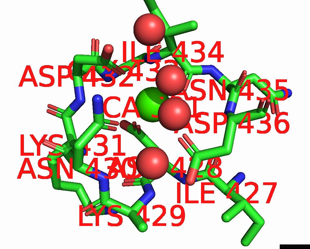
Mono view
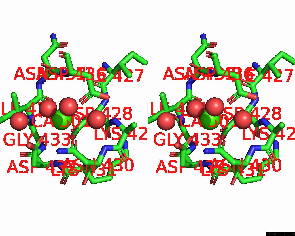
Stereo pair view
Mono view
Stereo pair view
A full contact list of Calcium with other atoms in the Ca binding
site number 1 of Structure of PFPL1 From Pseudoalteromonas Fuliginea within 5.0Å range:
|
Reference:
J.K.Hobbs,
A.B.Boraston.
The Structure of A Pectin-Active Family 1 Polysaccharide Lyase From the Marine Bacterium Pseudoalteromonas Fuliginea. Acta Crystallogr.,Sect.F 2024.
ISSN: ESSN 2053-230X
PubMed: 38935515
DOI: 10.1107/S2053230X2400596X
Page generated: Thu Jul 10 09:03:12 2025
ISSN: ESSN 2053-230X
PubMed: 38935515
DOI: 10.1107/S2053230X2400596X
Last articles
Cl in 5R9PCl in 5R9Q
Cl in 5R9O
Cl in 5R9N
Cl in 5R9M
Cl in 5R9K
Cl in 5R9L
Cl in 5R9J
Cl in 5R9I
Cl in 5R9G