Calcium »
PDB 9bi0-9ctr »
9c7i »
Calcium in PDB 9c7i: Crystal Structure of Caryolan-1-Ol Synthase From S. Griseus with Peg Molecule in the Active Site
Enzymatic activity of Crystal Structure of Caryolan-1-Ol Synthase From S. Griseus with Peg Molecule in the Active Site
All present enzymatic activity of Crystal Structure of Caryolan-1-Ol Synthase From S. Griseus with Peg Molecule in the Active Site:
4.2.1.138; 4.2.3.89;
4.2.1.138; 4.2.3.89;
Protein crystallography data
The structure of Crystal Structure of Caryolan-1-Ol Synthase From S. Griseus with Peg Molecule in the Active Site, PDB code: 9c7i
was solved by
R.Prem Kumar,
J.O.Matos,
D.D.Oprian,
with X-Ray Crystallography technique. A brief refinement statistics is given in the table below:
| Resolution Low / High (Å) | 70.57 / 2.33 |
| Space group | P 1 21 1 |
| Cell size a, b, c (Å), α, β, γ (°) | 68.522, 89.804, 114.11, 90, 90.82, 90 |
| R / Rfree (%) | 21.1 / 25.6 |
Calcium Binding Sites:
The binding sites of Calcium atom in the Crystal Structure of Caryolan-1-Ol Synthase From S. Griseus with Peg Molecule in the Active Site
(pdb code 9c7i). This binding sites where shown within
5.0 Angstroms radius around Calcium atom.
In total 6 binding sites of Calcium where determined in the Crystal Structure of Caryolan-1-Ol Synthase From S. Griseus with Peg Molecule in the Active Site, PDB code: 9c7i:
Jump to Calcium binding site number: 1; 2; 3; 4; 5; 6;
In total 6 binding sites of Calcium where determined in the Crystal Structure of Caryolan-1-Ol Synthase From S. Griseus with Peg Molecule in the Active Site, PDB code: 9c7i:
Jump to Calcium binding site number: 1; 2; 3; 4; 5; 6;
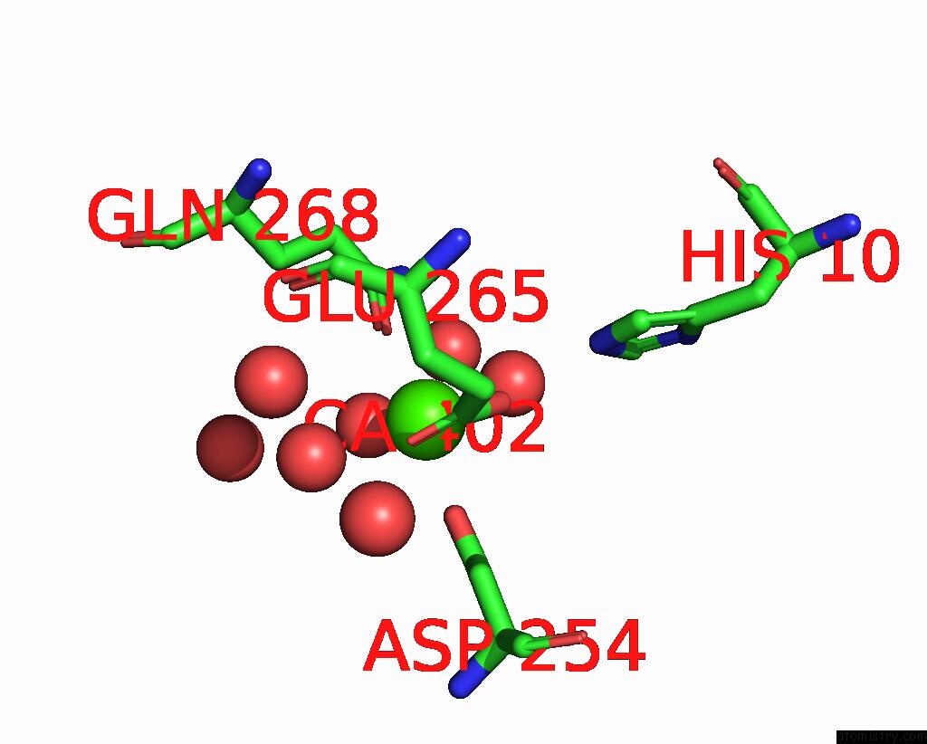
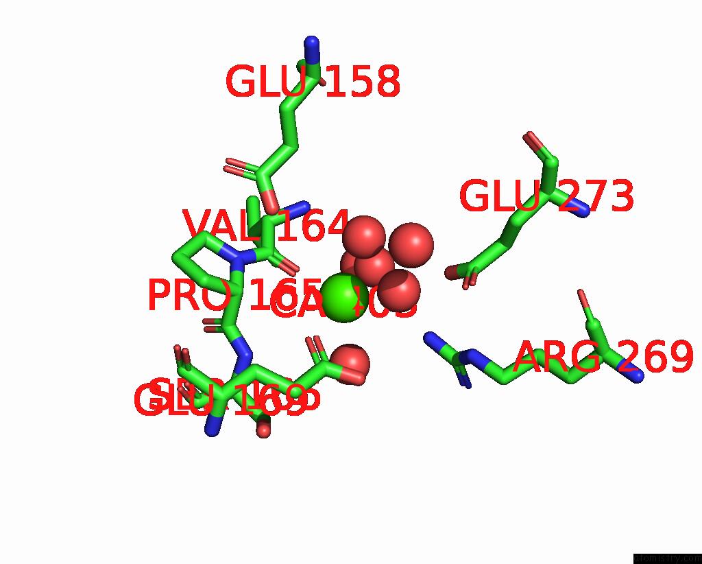
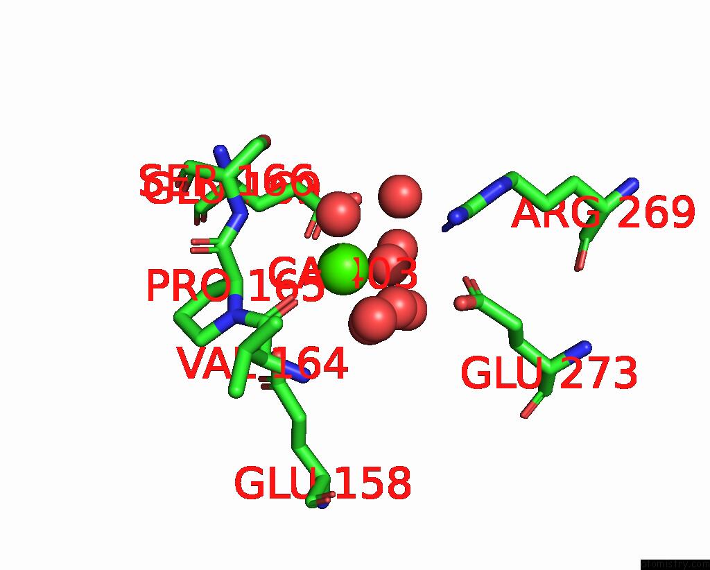
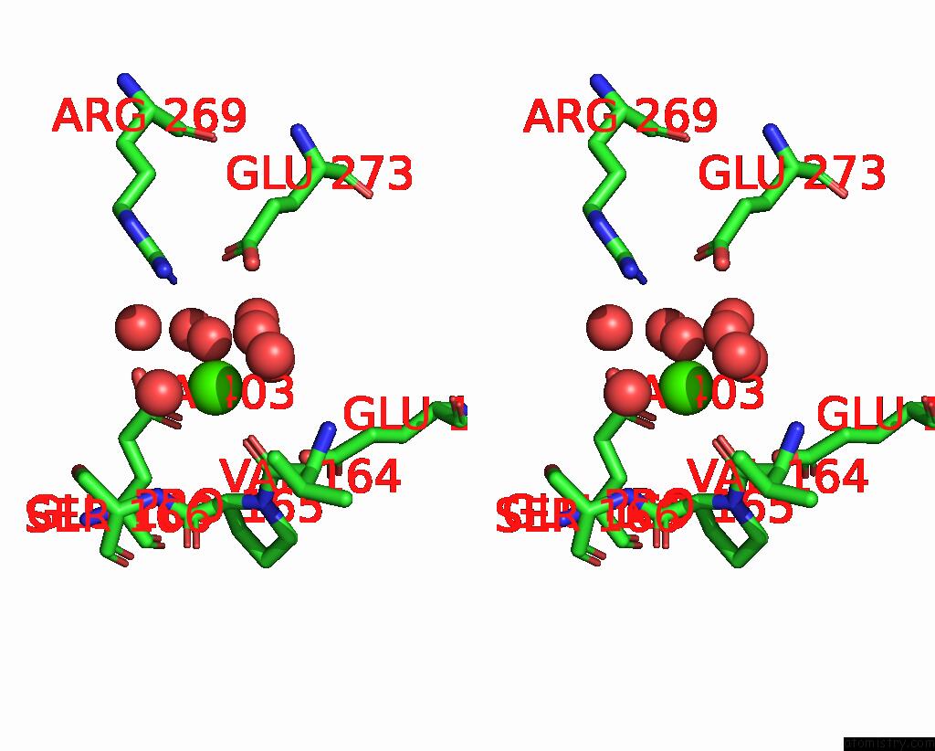
Calcium binding site 1 out of 6 in 9c7i
Go back to
Calcium binding site 1 out
of 6 in the Crystal Structure of Caryolan-1-Ol Synthase From S. Griseus with Peg Molecule in the Active Site
Mono view
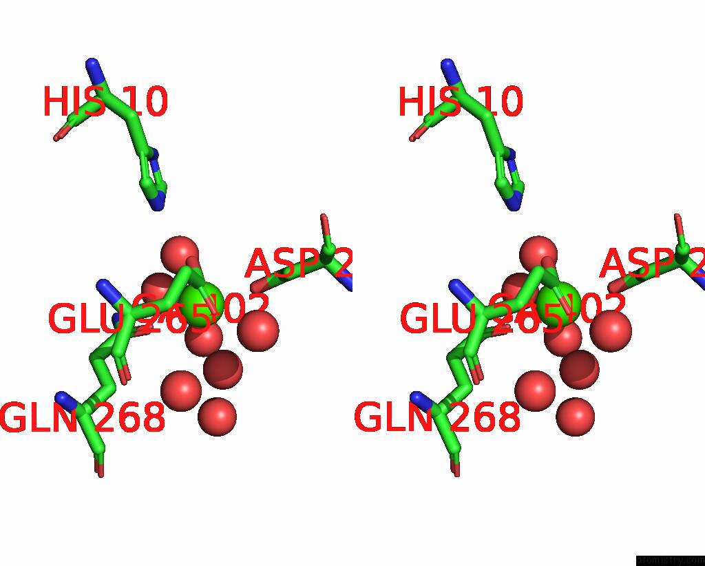
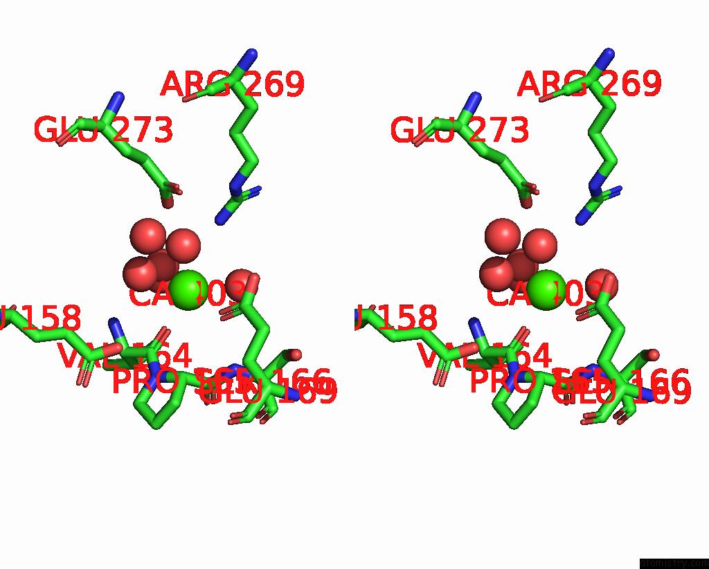
Stereo pair view
Mono view
Stereo pair view
A full contact list of Calcium with other atoms in the Ca binding
site number 1 of Crystal Structure of Caryolan-1-Ol Synthase From S. Griseus with Peg Molecule in the Active Site within 5.0Å range:
|
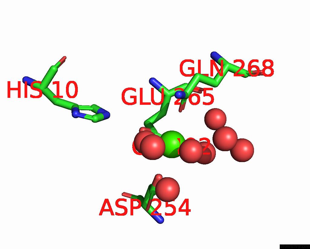
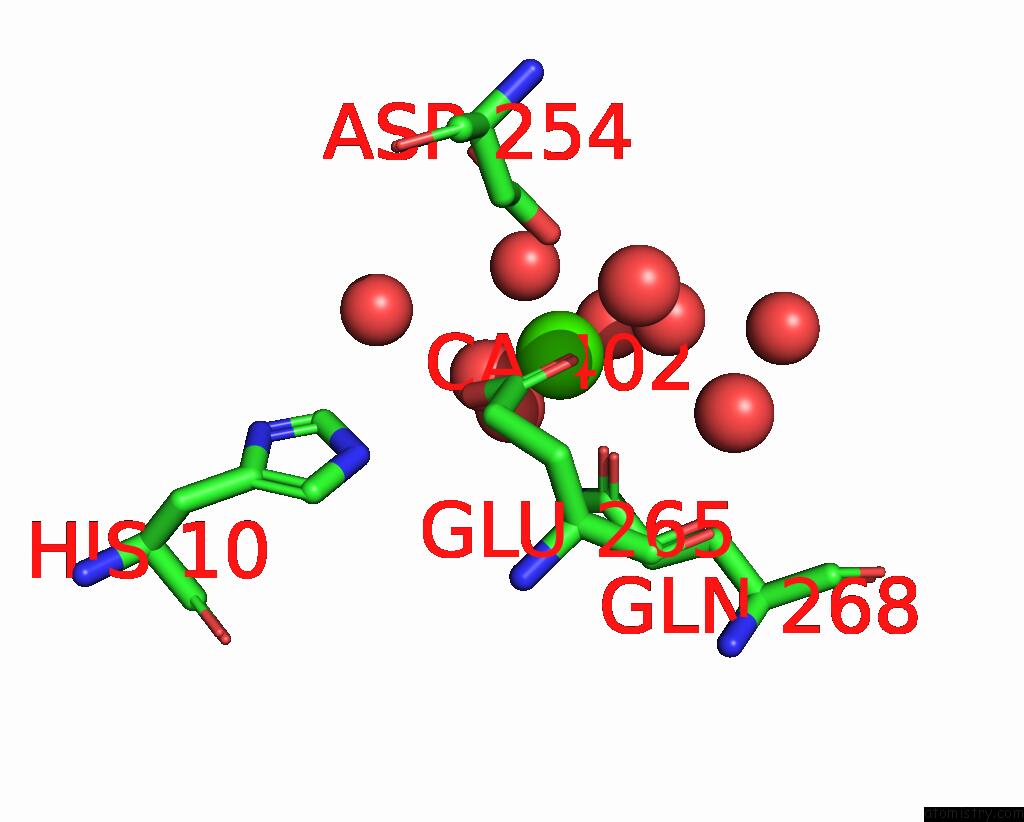
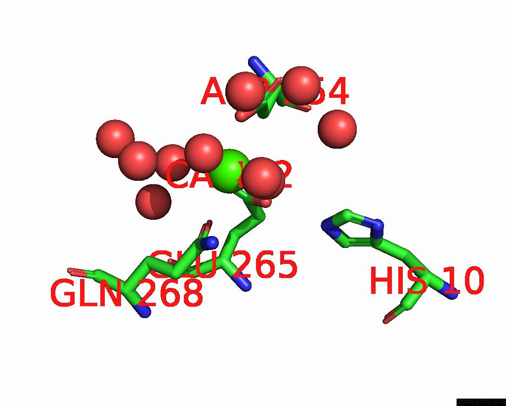
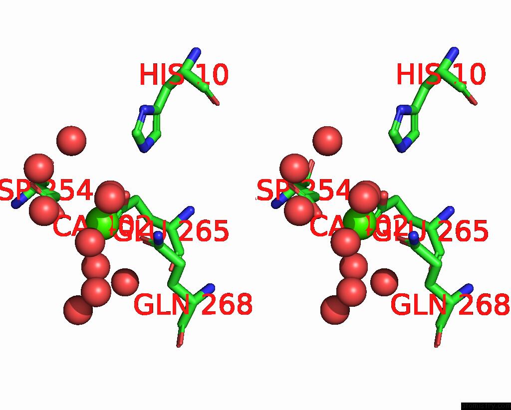
Calcium binding site 2 out of 6 in 9c7i
Go back to
Calcium binding site 2 out
of 6 in the Crystal Structure of Caryolan-1-Ol Synthase From S. Griseus with Peg Molecule in the Active Site
Mono view
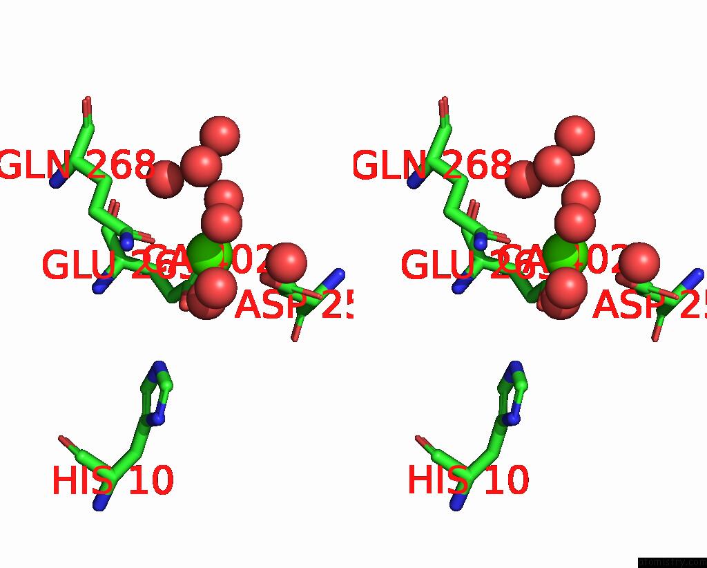
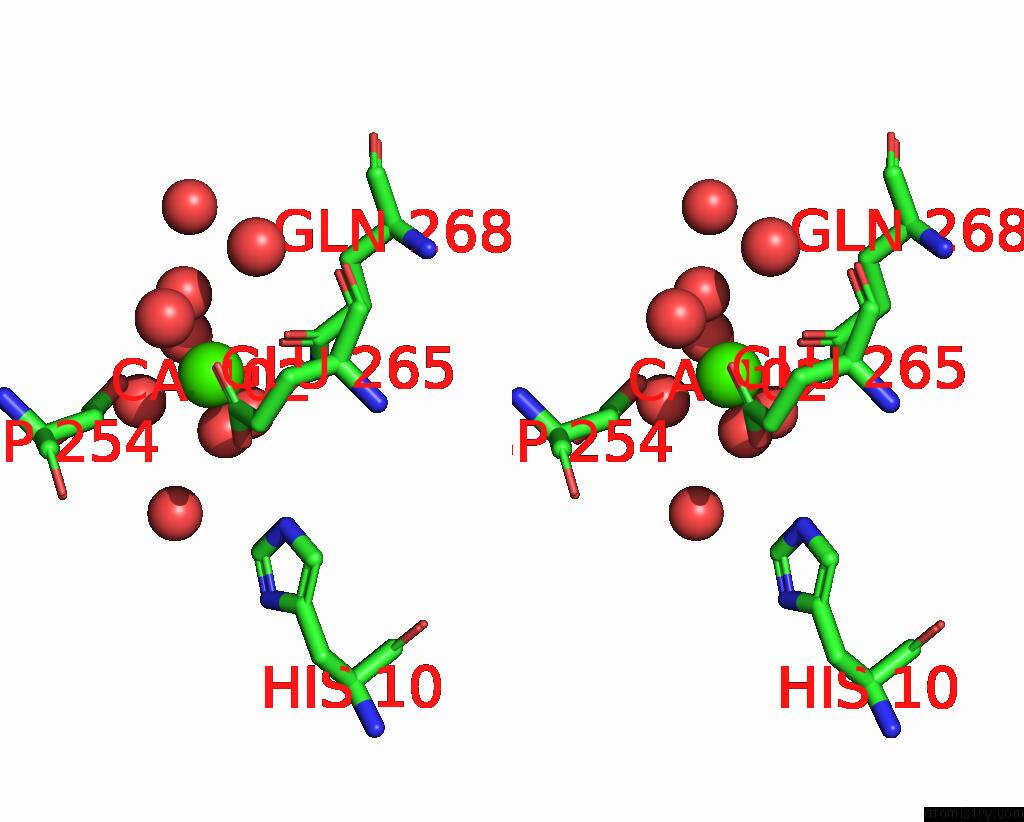
Stereo pair view
Mono view
Stereo pair view
A full contact list of Calcium with other atoms in the Ca binding
site number 2 of Crystal Structure of Caryolan-1-Ol Synthase From S. Griseus with Peg Molecule in the Active Site within 5.0Å range:
|
Calcium binding site 3 out of 6 in 9c7i
Go back to
Calcium binding site 3 out
of 6 in the Crystal Structure of Caryolan-1-Ol Synthase From S. Griseus with Peg Molecule in the Active Site
Mono view
Stereo pair view
Mono view
Stereo pair view
A full contact list of Calcium with other atoms in the Ca binding
site number 3 of Crystal Structure of Caryolan-1-Ol Synthase From S. Griseus with Peg Molecule in the Active Site within 5.0Å range:
|
Calcium binding site 4 out of 6 in 9c7i
Go back to
Calcium binding site 4 out
of 6 in the Crystal Structure of Caryolan-1-Ol Synthase From S. Griseus with Peg Molecule in the Active Site
Mono view
Stereo pair view
Mono view
Stereo pair view
A full contact list of Calcium with other atoms in the Ca binding
site number 4 of Crystal Structure of Caryolan-1-Ol Synthase From S. Griseus with Peg Molecule in the Active Site within 5.0Å range:
|
Calcium binding site 5 out of 6 in 9c7i
Go back to
Calcium binding site 5 out
of 6 in the Crystal Structure of Caryolan-1-Ol Synthase From S. Griseus with Peg Molecule in the Active Site
Mono view
Stereo pair view
Mono view
Stereo pair view
A full contact list of Calcium with other atoms in the Ca binding
site number 5 of Crystal Structure of Caryolan-1-Ol Synthase From S. Griseus with Peg Molecule in the Active Site within 5.0Å range:
|
Calcium binding site 6 out of 6 in 9c7i
Go back to
Calcium binding site 6 out
of 6 in the Crystal Structure of Caryolan-1-Ol Synthase From S. Griseus with Peg Molecule in the Active Site
Mono view
Stereo pair view
Mono view
Stereo pair view
A full contact list of Calcium with other atoms in the Ca binding
site number 6 of Crystal Structure of Caryolan-1-Ol Synthase From S. Griseus with Peg Molecule in the Active Site within 5.0Å range:
|
Reference:
R.P.Kumar,
J.O.Matos,
B.Y.Black,
W.H.Ellenburg,
J.Chen,
M.Patterson,
J.A.Gehtman,
D.L.Theobald,
I.J.Krauss,
D.D.Oprian.
Crystal Structure of Caryolan-1-Ol Synthase, A Sesquiterpene Synthase Catalyzing An Initial Anti-Markovnikov Cyclization Reaction. Biochemistry 2024.
ISSN: ISSN 0006-2960
PubMed: 39400323
DOI: 10.1021/ACS.BIOCHEM.4C00547
Page generated: Thu Jul 10 09:04:06 2025
ISSN: ISSN 0006-2960
PubMed: 39400323
DOI: 10.1021/ACS.BIOCHEM.4C00547
Last articles
Fe in 2YXOFe in 2YRS
Fe in 2YXC
Fe in 2YNM
Fe in 2YVJ
Fe in 2YP1
Fe in 2YU2
Fe in 2YU1
Fe in 2YQB
Fe in 2YOO