Calcium »
PDB 9bi0-9ctr »
9clp »
Calcium in PDB 9clp: Structure of Ecarin From the Venom of Kenyan Saw-Scaled Viper in Complex with the Fab of Neutralizing Antibody H11
Other elements in 9clp:
The structure of Structure of Ecarin From the Venom of Kenyan Saw-Scaled Viper in Complex with the Fab of Neutralizing Antibody H11 also contains other interesting chemical elements:
| Zinc | (Zn) | 1 atom |
Calcium Binding Sites:
The binding sites of Calcium atom in the Structure of Ecarin From the Venom of Kenyan Saw-Scaled Viper in Complex with the Fab of Neutralizing Antibody H11
(pdb code 9clp). This binding sites where shown within
5.0 Angstroms radius around Calcium atom.
In total 3 binding sites of Calcium where determined in the Structure of Ecarin From the Venom of Kenyan Saw-Scaled Viper in Complex with the Fab of Neutralizing Antibody H11, PDB code: 9clp:
Jump to Calcium binding site number: 1; 2; 3;
In total 3 binding sites of Calcium where determined in the Structure of Ecarin From the Venom of Kenyan Saw-Scaled Viper in Complex with the Fab of Neutralizing Antibody H11, PDB code: 9clp:
Jump to Calcium binding site number: 1; 2; 3;
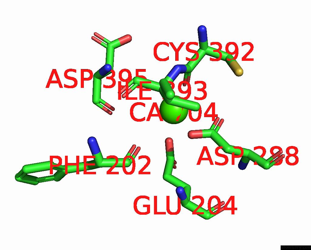
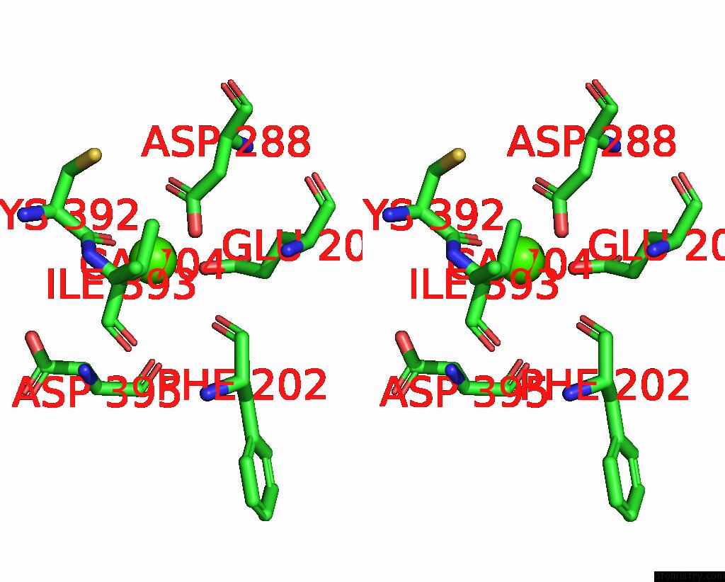
Calcium binding site 1 out of 3 in 9clp
Go back to
Calcium binding site 1 out
of 3 in the Structure of Ecarin From the Venom of Kenyan Saw-Scaled Viper in Complex with the Fab of Neutralizing Antibody H11
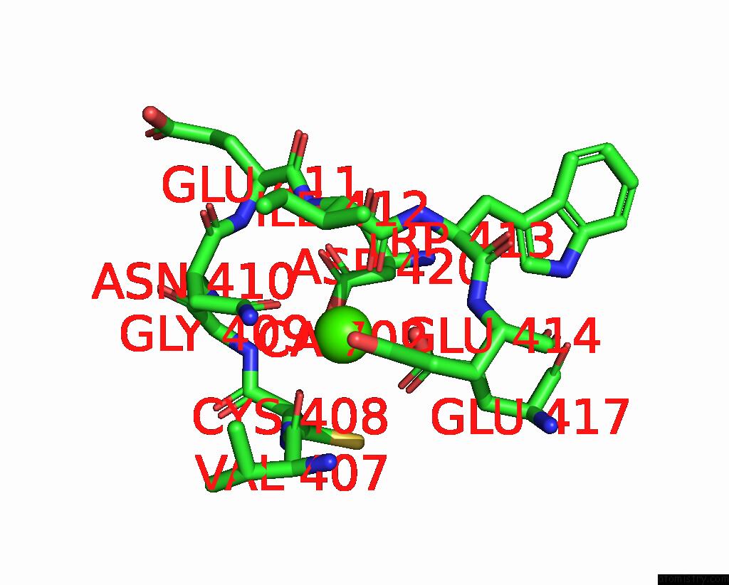
Mono view
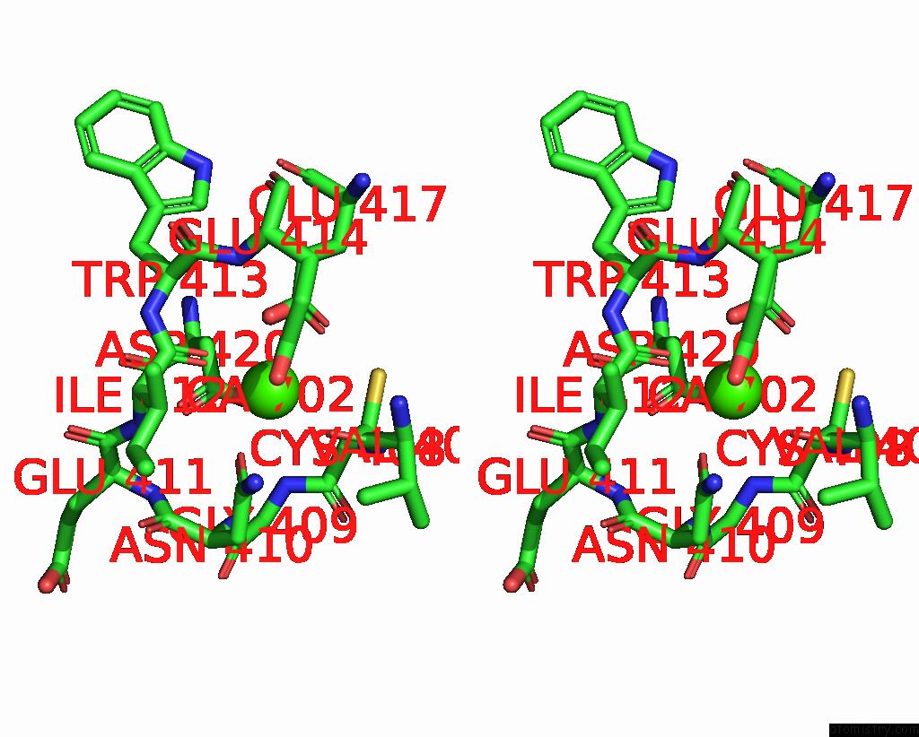
Stereo pair view
Mono view
Stereo pair view
A full contact list of Calcium with other atoms in the Ca binding
site number 1 of Structure of Ecarin From the Venom of Kenyan Saw-Scaled Viper in Complex with the Fab of Neutralizing Antibody H11 within 5.0Å range:
|
Calcium binding site 2 out of 3 in 9clp
Go back to
Calcium binding site 2 out
of 3 in the Structure of Ecarin From the Venom of Kenyan Saw-Scaled Viper in Complex with the Fab of Neutralizing Antibody H11
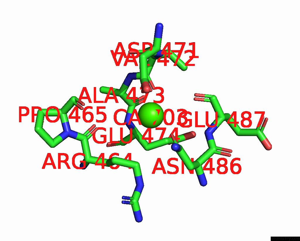
Mono view
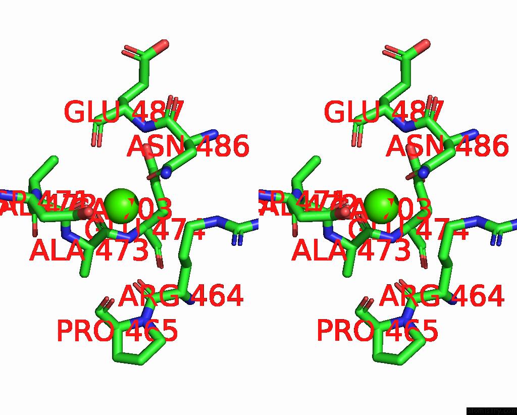
Stereo pair view
Mono view
Stereo pair view
A full contact list of Calcium with other atoms in the Ca binding
site number 2 of Structure of Ecarin From the Venom of Kenyan Saw-Scaled Viper in Complex with the Fab of Neutralizing Antibody H11 within 5.0Å range:
|
Calcium binding site 3 out of 3 in 9clp
Go back to
Calcium binding site 3 out
of 3 in the Structure of Ecarin From the Venom of Kenyan Saw-Scaled Viper in Complex with the Fab of Neutralizing Antibody H11
Mono view
Stereo pair view
Mono view
Stereo pair view
A full contact list of Calcium with other atoms in the Ca binding
site number 3 of Structure of Ecarin From the Venom of Kenyan Saw-Scaled Viper in Complex with the Fab of Neutralizing Antibody H11 within 5.0Å range:
|
Reference:
L.E.Misson Mindrebo,
J.T.Mindrebo,
Q.Tran,
M.C.Wilkinson,
J.M.Smith,
M.Verma,
N.R.Casewell,
G.C.Lander,
J.G.Jardine.
Importance of the Cysteine-Rich Domain of Snake Venom Prothrombin Activators: Insights Gained From Synthetic Neutralizing Antibodies. Toxins V. 16 2024.
ISSN: ESSN 2072-6651
PubMed: 39195771
DOI: 10.3390/TOXINS16080361
Page generated: Thu Jul 10 09:05:25 2025
ISSN: ESSN 2072-6651
PubMed: 39195771
DOI: 10.3390/TOXINS16080361
Last articles
Fe in 2YXOFe in 2YRS
Fe in 2YXC
Fe in 2YNM
Fe in 2YVJ
Fe in 2YP1
Fe in 2YU2
Fe in 2YU1
Fe in 2YQB
Fe in 2YOO