Calcium in PDB 9f8x: Low-Dose Structure of Marinobacter Nauticus Nitrous Oxide Reductase
Enzymatic activity of Low-Dose Structure of Marinobacter Nauticus Nitrous Oxide Reductase
All present enzymatic activity of Low-Dose Structure of Marinobacter Nauticus Nitrous Oxide Reductase:
1.7.2.4;
Protein crystallography data
The structure of Low-Dose Structure of Marinobacter Nauticus Nitrous Oxide Reductase, PDB code: 9f8x
was solved by
O.Einsle,
A.Pomowski,
with X-Ray Crystallography technique. A brief refinement statistics is given in the table below:
Resolution Low / High (Å)
|
23.50 /
1.50
|
Space group
|
P 1
|
Cell size a, b, c (Å), α, β, γ (°)
|
65.26,
69.375,
153.188,
81.38,
77.92,
88.61
|
R / Rfree (%)
|
13 /
15.2
|
Other elements in 9f8x:
The structure of Low-Dose Structure of Marinobacter Nauticus Nitrous Oxide Reductase also contains other interesting chemical elements:
Calcium Binding Sites:
The binding sites of Calcium atom in the Low-Dose Structure of Marinobacter Nauticus Nitrous Oxide Reductase
(pdb code 9f8x). This binding sites where shown within
5.0 Angstroms radius around Calcium atom.
In total 8 binding sites of Calcium where determined in the
Low-Dose Structure of Marinobacter Nauticus Nitrous Oxide Reductase, PDB code: 9f8x:
Jump to Calcium binding site number:
1;
2;
3;
4;
5;
6;
7;
8;
Calcium binding site 1 out
of 8 in 9f8x
Go back to
Calcium Binding Sites List in 9f8x
Calcium binding site 1 out
of 8 in the Low-Dose Structure of Marinobacter Nauticus Nitrous Oxide Reductase
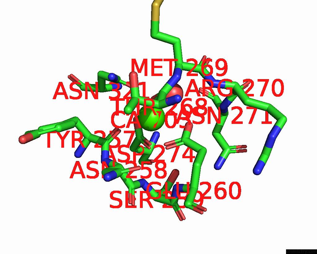
Mono view
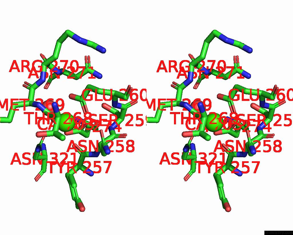
Stereo pair view
|
A full contact list of Calcium with other atoms in the Ca binding
site number 1 of Low-Dose Structure of Marinobacter Nauticus Nitrous Oxide Reductase within 5.0Å range:
probe
|
atom
|
residue
|
distance (Å)
|
B
|
Occ
|
A:Ca703
b:19.4
occ:1.00
|
O
|
A:TYR257
|
2.4
|
18.1
|
1.0
|
O
|
A:THR268
|
2.4
|
22.3
|
1.0
|
OD1
|
A:ASN321
|
2.4
|
18.6
|
1.0
|
O
|
A:HOH820
|
2.5
|
22.2
|
1.0
|
OD1
|
A:ASP274
|
2.5
|
20.3
|
1.0
|
OE1
|
A:GLU260
|
2.6
|
21.4
|
1.0
|
OE2
|
A:GLU260
|
2.6
|
22.2
|
1.0
|
OD2
|
A:ASP274
|
2.7
|
19.3
|
1.0
|
CD
|
A:GLU260
|
2.9
|
22.1
|
1.0
|
CG
|
A:ASP274
|
2.9
|
18.6
|
1.0
|
CG
|
A:ASN321
|
3.4
|
17.7
|
1.0
|
C
|
A:TYR257
|
3.5
|
17.5
|
1.0
|
C
|
A:THR268
|
3.5
|
23.4
|
1.0
|
ND2
|
A:ASN321
|
3.9
|
17.9
|
1.0
|
CA
|
A:MET269
|
4.2
|
26.7
|
1.0
|
N
|
A:MET269
|
4.3
|
24.5
|
1.0
|
N
|
A:ASN258
|
4.3
|
18.0
|
1.0
|
CA
|
A:ASN258
|
4.3
|
19.5
|
1.0
|
OG
|
A:SER259
|
4.4
|
22.9
|
1.0
|
N
|
A:SER259
|
4.4
|
20.0
|
1.0
|
CA
|
A:TYR257
|
4.4
|
16.5
|
1.0
|
CG
|
A:GLU260
|
4.4
|
21.0
|
1.0
|
CB
|
A:ASP274
|
4.4
|
17.8
|
1.0
|
N
|
A:ASN321
|
4.5
|
16.2
|
1.0
|
CA
|
A:THR268
|
4.5
|
23.0
|
1.0
|
CA
|
A:ASN321
|
4.5
|
15.3
|
1.0
|
CB
|
A:ASN321
|
4.6
|
15.8
|
1.0
|
C
|
A:MET269
|
4.7
|
26.5
|
1.0
|
O
|
A:ASN271
|
4.8
|
24.4
|
1.0
|
C
|
A:ASN258
|
4.9
|
20.1
|
1.0
|
N
|
A:ARG270
|
4.9
|
24.5
|
1.0
|
N
|
A:ASN271
|
4.9
|
23.7
|
1.0
|
|
Calcium binding site 2 out
of 8 in 9f8x
Go back to
Calcium Binding Sites List in 9f8x
Calcium binding site 2 out
of 8 in the Low-Dose Structure of Marinobacter Nauticus Nitrous Oxide Reductase
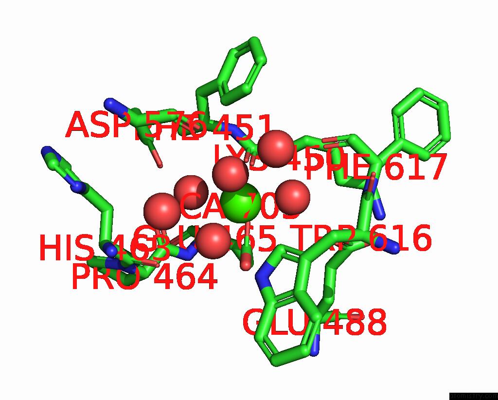
Mono view
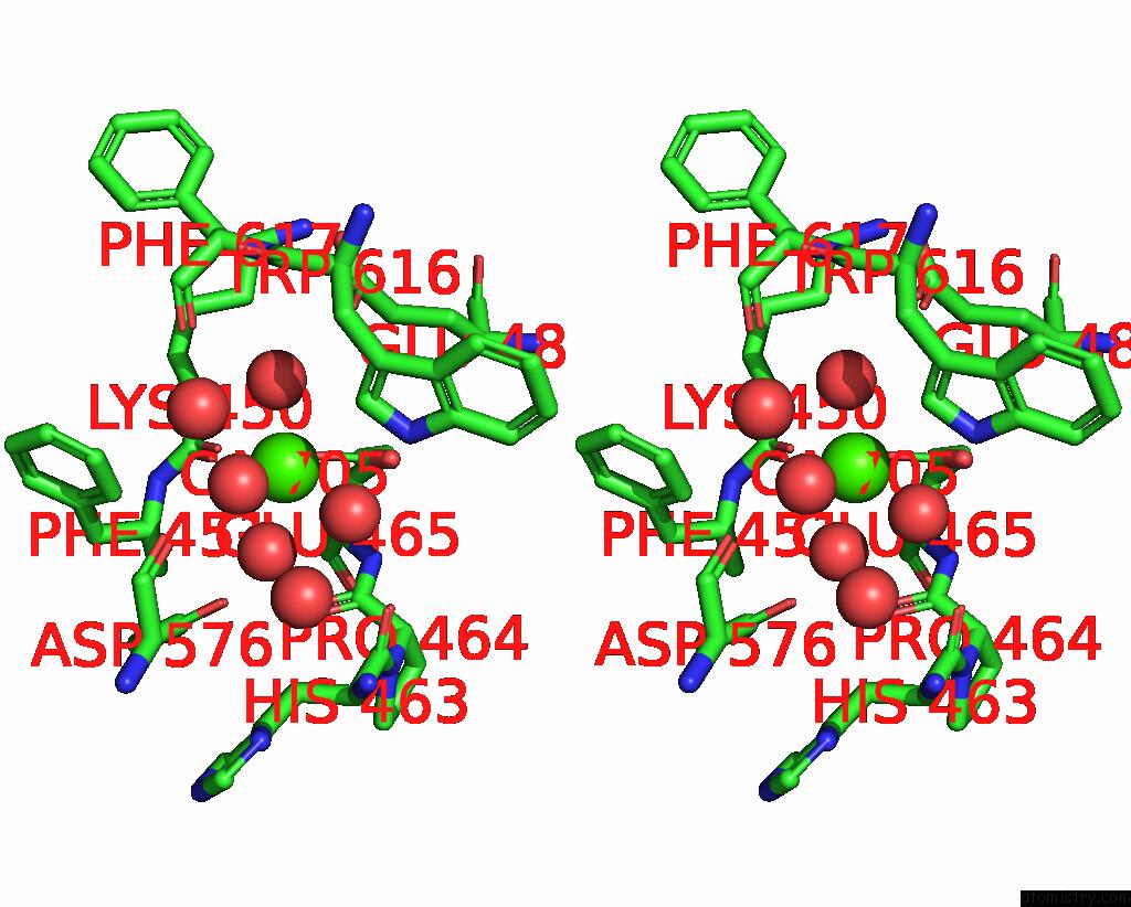
Stereo pair view
|
A full contact list of Calcium with other atoms in the Ca binding
site number 2 of Low-Dose Structure of Marinobacter Nauticus Nitrous Oxide Reductase within 5.0Å range:
probe
|
atom
|
residue
|
distance (Å)
|
B
|
Occ
|
A:Ca705
b:16.9
occ:1.00
|
OE1
|
A:GLU465
|
2.2
|
27.5
|
1.0
|
O
|
A:LYS450
|
2.2
|
16.7
|
1.0
|
O
|
B:HOH921
|
2.3
|
15.0
|
1.0
|
O
|
B:HOH1135
|
2.3
|
16.3
|
1.0
|
O
|
A:HOH1132
|
2.4
|
15.4
|
1.0
|
O
|
A:HOH1246
|
2.5
|
20.4
|
1.0
|
CD
|
A:GLU465
|
2.9
|
25.6
|
1.0
|
OE2
|
A:GLU465
|
3.0
|
30.4
|
1.0
|
C
|
A:LYS450
|
3.4
|
17.2
|
1.0
|
OD1
|
B:ASP576
|
4.1
|
17.4
|
1.0
|
CA
|
A:LYS450
|
4.2
|
16.1
|
1.0
|
CD1
|
B:TRP616
|
4.3
|
15.1
|
1.0
|
N
|
A:PHE451
|
4.3
|
15.6
|
1.0
|
CG
|
A:GLU465
|
4.4
|
21.1
|
1.0
|
CA
|
A:PHE451
|
4.5
|
16.0
|
1.0
|
O
|
B:HOH949
|
4.5
|
16.3
|
1.0
|
NE1
|
B:TRP616
|
4.5
|
15.8
|
1.0
|
CD1
|
A:PHE451
|
4.5
|
18.5
|
1.0
|
O
|
A:HIS463
|
4.6
|
16.4
|
1.0
|
O
|
B:HOH979
|
4.6
|
16.5
|
1.0
|
OE1
|
A:GLU488
|
4.6
|
19.6
|
1.0
|
O
|
A:PRO464
|
4.7
|
14.1
|
1.0
|
O
|
B:PHE617
|
4.7
|
16.1
|
1.0
|
CB
|
A:LYS450
|
4.8
|
15.7
|
1.0
|
CA
|
A:GLU465
|
4.9
|
15.4
|
1.0
|
CG
|
B:ASP576
|
5.0
|
16.6
|
1.0
|
CB
|
A:GLU465
|
5.0
|
17.5
|
1.0
|
C
|
A:PRO464
|
5.0
|
14.2
|
1.0
|
|
Calcium binding site 3 out
of 8 in 9f8x
Go back to
Calcium Binding Sites List in 9f8x
Calcium binding site 3 out
of 8 in the Low-Dose Structure of Marinobacter Nauticus Nitrous Oxide Reductase
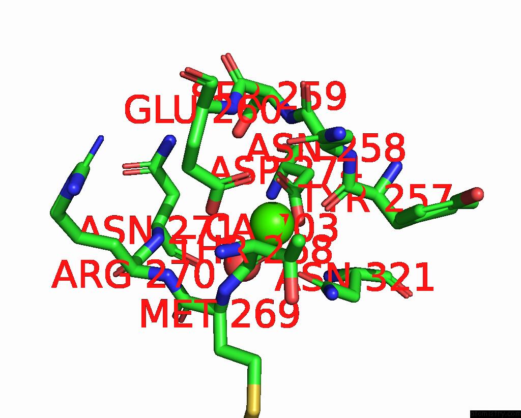
Mono view
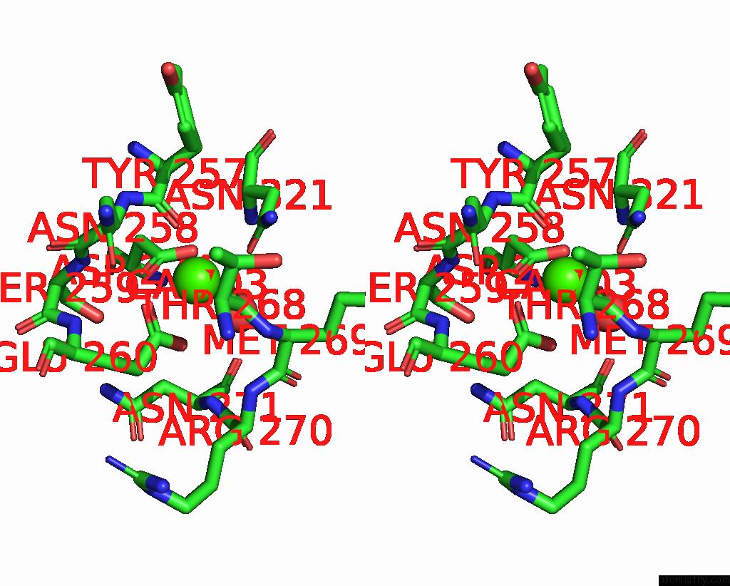
Stereo pair view
|
A full contact list of Calcium with other atoms in the Ca binding
site number 3 of Low-Dose Structure of Marinobacter Nauticus Nitrous Oxide Reductase within 5.0Å range:
probe
|
atom
|
residue
|
distance (Å)
|
B
|
Occ
|
B:Ca703
b:13.6
occ:1.00
|
O
|
B:TYR257
|
2.4
|
14.3
|
1.0
|
O
|
B:HOH870
|
2.4
|
16.2
|
1.0
|
O
|
B:THR268
|
2.4
|
15.3
|
1.0
|
OD1
|
B:ASN321
|
2.4
|
15.2
|
1.0
|
OD1
|
B:ASP274
|
2.5
|
14.4
|
1.0
|
OE2
|
B:GLU260
|
2.5
|
15.4
|
1.0
|
OE1
|
B:GLU260
|
2.5
|
15.6
|
1.0
|
OD2
|
B:ASP274
|
2.6
|
15.2
|
1.0
|
CD
|
B:GLU260
|
2.9
|
15.4
|
1.0
|
CG
|
B:ASP274
|
2.9
|
14.4
|
1.0
|
CG
|
B:ASN321
|
3.4
|
14.0
|
1.0
|
C
|
B:TYR257
|
3.5
|
13.1
|
1.0
|
C
|
B:THR268
|
3.5
|
14.6
|
1.0
|
ND2
|
B:ASN321
|
3.9
|
13.9
|
1.0
|
N
|
B:ASN258
|
4.3
|
13.4
|
1.0
|
N
|
B:MET269
|
4.3
|
15.2
|
1.0
|
CA
|
B:MET269
|
4.3
|
16.2
|
1.0
|
OG
|
B:SER259
|
4.3
|
16.2
|
1.0
|
CA
|
B:ASN258
|
4.3
|
13.9
|
1.0
|
N
|
B:SER259
|
4.4
|
13.3
|
1.0
|
CG
|
B:GLU260
|
4.4
|
15.1
|
1.0
|
CA
|
B:TYR257
|
4.4
|
12.5
|
1.0
|
CB
|
B:ASP274
|
4.4
|
13.2
|
1.0
|
CA
|
B:THR268
|
4.5
|
15.2
|
1.0
|
N
|
B:ASN321
|
4.5
|
13.3
|
1.0
|
CB
|
B:ASN321
|
4.6
|
13.8
|
1.0
|
CA
|
B:ASN321
|
4.6
|
13.5
|
1.0
|
O
|
B:ASN271
|
4.7
|
16.3
|
1.0
|
C
|
B:MET269
|
4.7
|
16.8
|
1.0
|
N
|
B:ASN271
|
4.8
|
17.5
|
1.0
|
N
|
B:ARG270
|
4.8
|
16.6
|
1.0
|
C
|
B:ASN258
|
4.9
|
13.8
|
1.0
|
CB
|
B:ASN271
|
5.0
|
16.6
|
1.0
|
|
Calcium binding site 4 out
of 8 in 9f8x
Go back to
Calcium Binding Sites List in 9f8x
Calcium binding site 4 out
of 8 in the Low-Dose Structure of Marinobacter Nauticus Nitrous Oxide Reductase
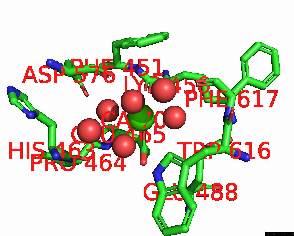
Mono view
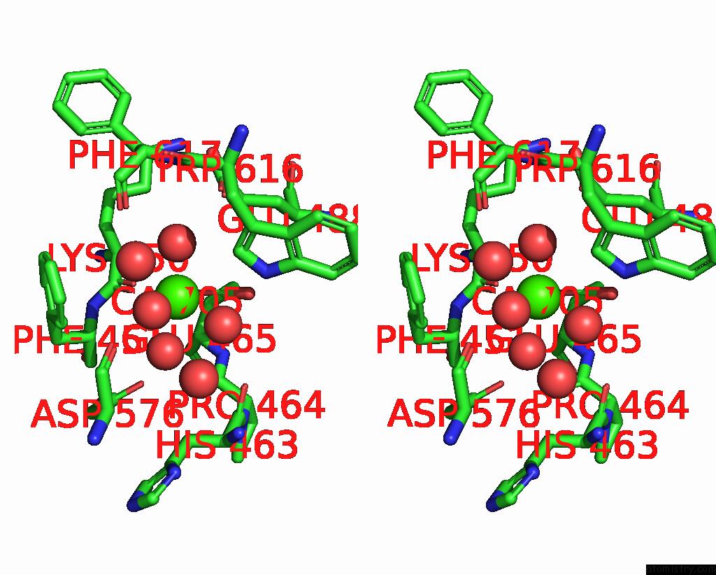
Stereo pair view
|
A full contact list of Calcium with other atoms in the Ca binding
site number 4 of Low-Dose Structure of Marinobacter Nauticus Nitrous Oxide Reductase within 5.0Å range:
probe
|
atom
|
residue
|
distance (Å)
|
B
|
Occ
|
B:Ca705
b:16.2
occ:1.00
|
O
|
B:LYS450
|
2.2
|
17.7
|
1.0
|
OE1
|
B:GLU465
|
2.2
|
27.9
|
1.0
|
O
|
B:HOH1186
|
2.3
|
17.0
|
1.0
|
O
|
A:HOH933
|
2.3
|
15.4
|
1.0
|
O
|
A:HOH1034
|
2.4
|
17.4
|
1.0
|
O
|
B:HOH1328
|
2.5
|
19.4
|
1.0
|
CD
|
B:GLU465
|
3.1
|
23.8
|
1.0
|
OE2
|
B:GLU465
|
3.3
|
27.7
|
1.0
|
C
|
B:LYS450
|
3.4
|
17.0
|
1.0
|
OD1
|
A:ASP576
|
4.1
|
17.2
|
1.0
|
CA
|
B:LYS450
|
4.2
|
16.1
|
1.0
|
N
|
B:PHE451
|
4.3
|
17.4
|
1.0
|
CA
|
B:PHE451
|
4.4
|
17.2
|
1.0
|
O
|
A:HOH988
|
4.4
|
15.7
|
1.0
|
CD1
|
A:TRP616
|
4.4
|
14.4
|
1.0
|
CG
|
B:GLU465
|
4.5
|
20.0
|
1.0
|
O
|
A:HOH1009
|
4.6
|
16.3
|
1.0
|
O
|
B:HIS463
|
4.6
|
14.7
|
1.0
|
NE1
|
A:TRP616
|
4.6
|
14.6
|
1.0
|
O
|
A:PHE617
|
4.6
|
15.8
|
1.0
|
OE1
|
B:GLU488
|
4.7
|
20.2
|
1.0
|
O
|
B:PRO464
|
4.7
|
14.9
|
1.0
|
CB
|
B:LYS450
|
4.7
|
16.8
|
1.0
|
CA
|
B:GLU465
|
4.9
|
15.6
|
1.0
|
CD2
|
B:PHE451
|
4.9
|
24.4
|
1.0
|
CB
|
B:GLU465
|
4.9
|
17.1
|
1.0
|
C
|
B:PRO464
|
5.0
|
15.0
|
1.0
|
N
|
B:GLU465
|
5.0
|
14.1
|
1.0
|
|
Calcium binding site 5 out
of 8 in 9f8x
Go back to
Calcium Binding Sites List in 9f8x
Calcium binding site 5 out
of 8 in the Low-Dose Structure of Marinobacter Nauticus Nitrous Oxide Reductase
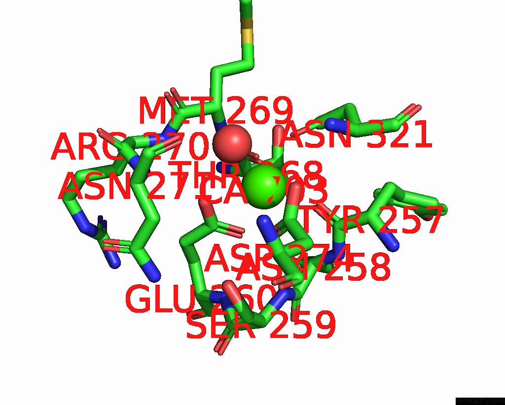
Mono view
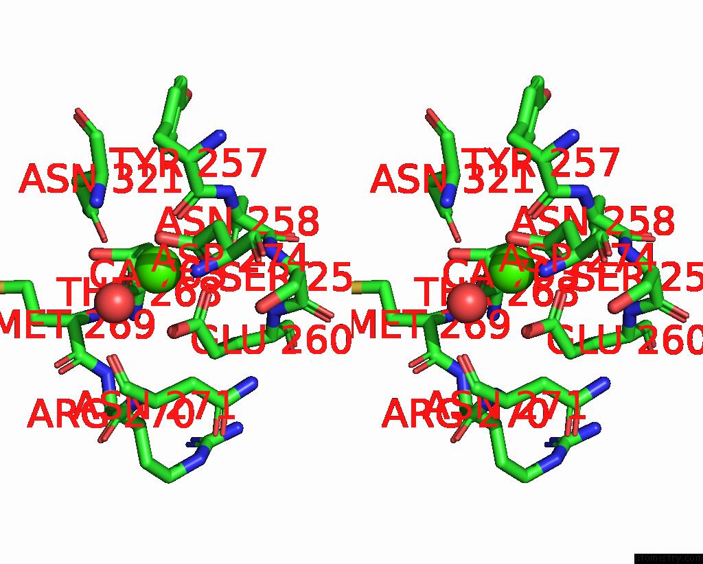
Stereo pair view
|
A full contact list of Calcium with other atoms in the Ca binding
site number 5 of Low-Dose Structure of Marinobacter Nauticus Nitrous Oxide Reductase within 5.0Å range:
probe
|
atom
|
residue
|
distance (Å)
|
B
|
Occ
|
C:Ca703
b:12.5
occ:1.00
|
O
|
C:THR268
|
2.3
|
13.6
|
1.0
|
O
|
C:TYR257
|
2.4
|
13.1
|
1.0
|
O
|
C:HOH880
|
2.4
|
15.4
|
1.0
|
OD1
|
C:ASN321
|
2.4
|
13.7
|
1.0
|
OD1
|
C:ASP274
|
2.5
|
12.9
|
1.0
|
OE2
|
C:GLU260
|
2.5
|
14.2
|
1.0
|
OE1
|
C:GLU260
|
2.5
|
13.8
|
1.0
|
OD2
|
C:ASP274
|
2.7
|
13.9
|
1.0
|
CD
|
C:GLU260
|
2.9
|
15.2
|
1.0
|
CG
|
C:ASP274
|
2.9
|
13.6
|
1.0
|
CG
|
C:ASN321
|
3.4
|
12.9
|
1.0
|
C
|
C:THR268
|
3.5
|
14.0
|
1.0
|
C
|
C:TYR257
|
3.5
|
12.4
|
1.0
|
ND2
|
C:ASN321
|
3.9
|
13.5
|
1.0
|
N
|
C:MET269
|
4.3
|
14.1
|
1.0
|
N
|
C:ASN258
|
4.3
|
13.1
|
1.0
|
CA
|
C:MET269
|
4.3
|
15.8
|
1.0
|
CG
|
C:GLU260
|
4.4
|
14.8
|
1.0
|
CA
|
C:ASN258
|
4.4
|
13.4
|
1.0
|
OG
|
C:SER259
|
4.4
|
14.8
|
1.0
|
CA
|
C:TYR257
|
4.4
|
12.3
|
1.0
|
CB
|
C:ASP274
|
4.4
|
13.2
|
1.0
|
CA
|
C:THR268
|
4.4
|
14.5
|
1.0
|
N
|
C:SER259
|
4.4
|
13.2
|
1.0
|
N
|
C:ASN321
|
4.6
|
12.8
|
1.0
|
CB
|
C:ASN321
|
4.6
|
11.9
|
1.0
|
CA
|
C:ASN321
|
4.6
|
12.4
|
1.0
|
C
|
C:MET269
|
4.7
|
15.2
|
1.0
|
O
|
C:ASN271
|
4.7
|
16.1
|
1.0
|
N
|
C:ARG270
|
4.8
|
15.3
|
1.0
|
N
|
C:ASN271
|
4.9
|
14.8
|
1.0
|
C
|
C:ASN258
|
4.9
|
13.3
|
1.0
|
|
Calcium binding site 6 out
of 8 in 9f8x
Go back to
Calcium Binding Sites List in 9f8x
Calcium binding site 6 out
of 8 in the Low-Dose Structure of Marinobacter Nauticus Nitrous Oxide Reductase
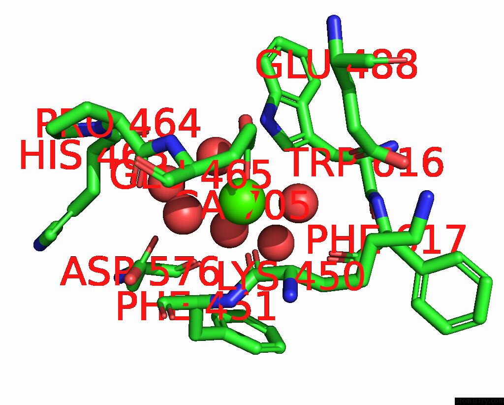
Mono view
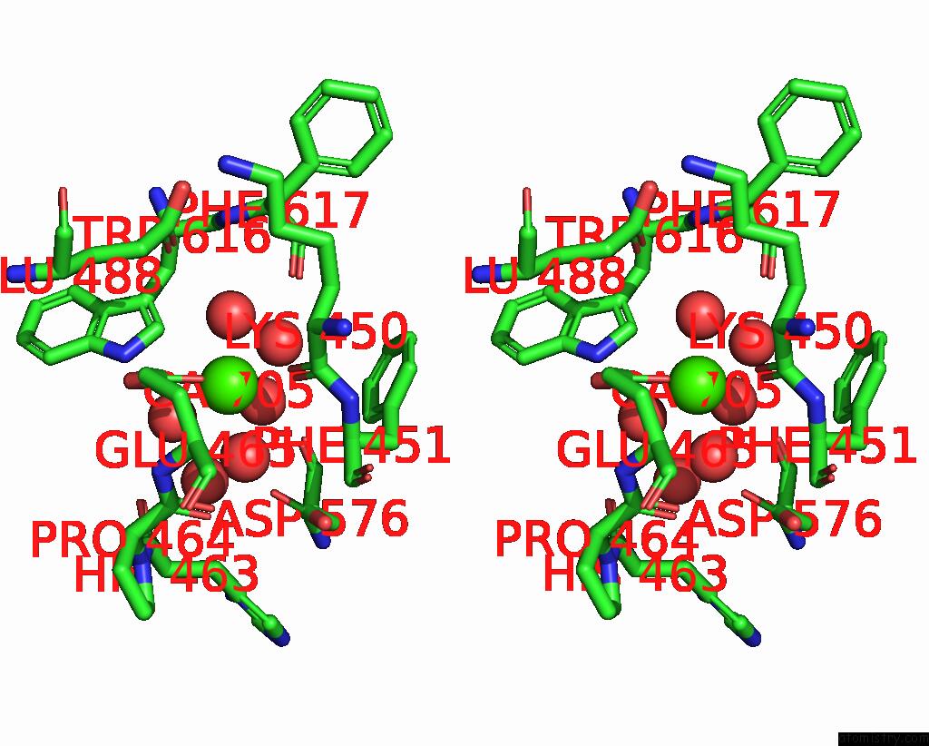
Stereo pair view
|
A full contact list of Calcium with other atoms in the Ca binding
site number 6 of Low-Dose Structure of Marinobacter Nauticus Nitrous Oxide Reductase within 5.0Å range:
probe
|
atom
|
residue
|
distance (Å)
|
B
|
Occ
|
C:Ca705
b:14.7
occ:1.00
|
OE1
|
C:GLU465
|
2.2
|
24.3
|
1.0
|
O
|
D:HOH1187
|
2.3
|
15.9
|
1.0
|
O
|
C:LYS450
|
2.3
|
17.2
|
1.0
|
O
|
D:HOH1005
|
2.3
|
13.3
|
1.0
|
O
|
D:HOH1015
|
2.4
|
15.7
|
1.0
|
O
|
D:HOH1307
|
2.4
|
18.2
|
1.0
|
CD
|
C:GLU465
|
3.1
|
23.8
|
1.0
|
OE2
|
C:GLU465
|
3.3
|
28.2
|
1.0
|
C
|
C:LYS450
|
3.5
|
15.8
|
1.0
|
OD1
|
D:ASP576
|
4.1
|
15.2
|
1.0
|
CA
|
C:LYS450
|
4.3
|
15.3
|
1.0
|
CD1
|
D:TRP616
|
4.4
|
14.2
|
1.0
|
N
|
C:PHE451
|
4.4
|
16.1
|
1.0
|
O
|
D:HOH953
|
4.4
|
14.2
|
1.0
|
CA
|
C:PHE451
|
4.4
|
15.4
|
1.0
|
CG
|
C:GLU465
|
4.5
|
19.3
|
1.0
|
O
|
D:HOH935
|
4.5
|
15.0
|
1.0
|
NE1
|
D:TRP616
|
4.5
|
13.8
|
1.0
|
O
|
C:HIS463
|
4.6
|
14.8
|
1.0
|
O
|
D:PHE617
|
4.6
|
14.2
|
1.0
|
O
|
C:PRO464
|
4.7
|
14.1
|
1.0
|
OE1
|
C:GLU488
|
4.7
|
19.8
|
1.0
|
CB
|
C:LYS450
|
4.8
|
15.2
|
1.0
|
CD1
|
C:PHE451
|
4.8
|
20.1
|
1.0
|
CA
|
C:GLU465
|
4.9
|
14.6
|
1.0
|
CB
|
C:GLU465
|
5.0
|
15.6
|
1.0
|
C
|
C:PRO464
|
5.0
|
13.8
|
1.0
|
|
Calcium binding site 7 out
of 8 in 9f8x
Go back to
Calcium Binding Sites List in 9f8x
Calcium binding site 7 out
of 8 in the Low-Dose Structure of Marinobacter Nauticus Nitrous Oxide Reductase
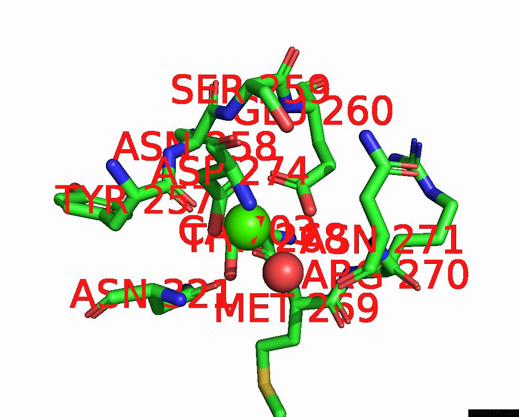
Mono view
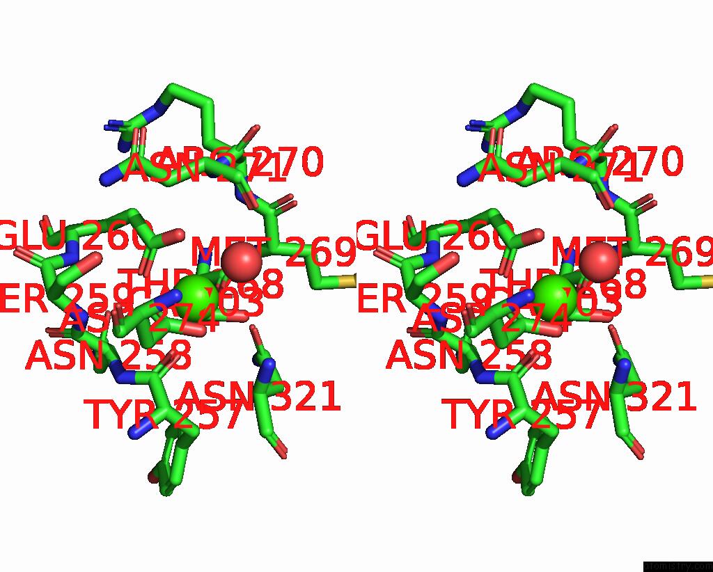
Stereo pair view
|
A full contact list of Calcium with other atoms in the Ca binding
site number 7 of Low-Dose Structure of Marinobacter Nauticus Nitrous Oxide Reductase within 5.0Å range:
probe
|
atom
|
residue
|
distance (Å)
|
B
|
Occ
|
D:Ca703
b:16.8
occ:1.00
|
O
|
D:TYR257
|
2.4
|
15.9
|
1.0
|
O
|
D:THR268
|
2.4
|
17.9
|
1.0
|
O
|
D:HOH843
|
2.4
|
18.9
|
1.0
|
OD1
|
D:ASP274
|
2.5
|
17.6
|
1.0
|
OD1
|
D:ASN321
|
2.5
|
15.9
|
1.0
|
OE2
|
D:GLU260
|
2.5
|
19.3
|
1.0
|
OE1
|
D:GLU260
|
2.6
|
19.3
|
1.0
|
OD2
|
D:ASP274
|
2.7
|
16.9
|
1.0
|
CD
|
D:GLU260
|
2.9
|
19.5
|
1.0
|
CG
|
D:ASP274
|
2.9
|
17.2
|
1.0
|
CG
|
D:ASN321
|
3.4
|
15.0
|
1.0
|
C
|
D:TYR257
|
3.5
|
15.2
|
1.0
|
C
|
D:THR268
|
3.5
|
20.4
|
1.0
|
ND2
|
D:ASN321
|
3.9
|
15.4
|
1.0
|
CA
|
D:MET269
|
4.2
|
23.4
|
1.0
|
N
|
D:ASN258
|
4.3
|
16.8
|
1.0
|
N
|
D:MET269
|
4.3
|
22.5
|
1.0
|
CA
|
D:ASN258
|
4.4
|
16.9
|
1.0
|
N
|
D:SER259
|
4.4
|
17.8
|
1.0
|
OG
|
D:SER259
|
4.4
|
18.9
|
1.0
|
CG
|
D:GLU260
|
4.4
|
19.9
|
1.0
|
CA
|
D:TYR257
|
4.4
|
15.3
|
1.0
|
CB
|
D:ASP274
|
4.4
|
15.6
|
1.0
|
CA
|
D:THR268
|
4.5
|
19.3
|
1.0
|
N
|
D:ASN321
|
4.5
|
13.9
|
1.0
|
CA
|
D:ASN321
|
4.6
|
14.9
|
1.0
|
CB
|
D:ASN321
|
4.6
|
14.1
|
1.0
|
C
|
D:MET269
|
4.7
|
26.0
|
1.0
|
O
|
D:ASN271
|
4.8
|
22.6
|
1.0
|
N
|
D:ARG270
|
4.8
|
22.2
|
1.0
|
N
|
D:ASN271
|
4.9
|
21.9
|
1.0
|
C
|
D:ASN258
|
4.9
|
17.9
|
1.0
|
|
Calcium binding site 8 out
of 8 in 9f8x
Go back to
Calcium Binding Sites List in 9f8x
Calcium binding site 8 out
of 8 in the Low-Dose Structure of Marinobacter Nauticus Nitrous Oxide Reductase
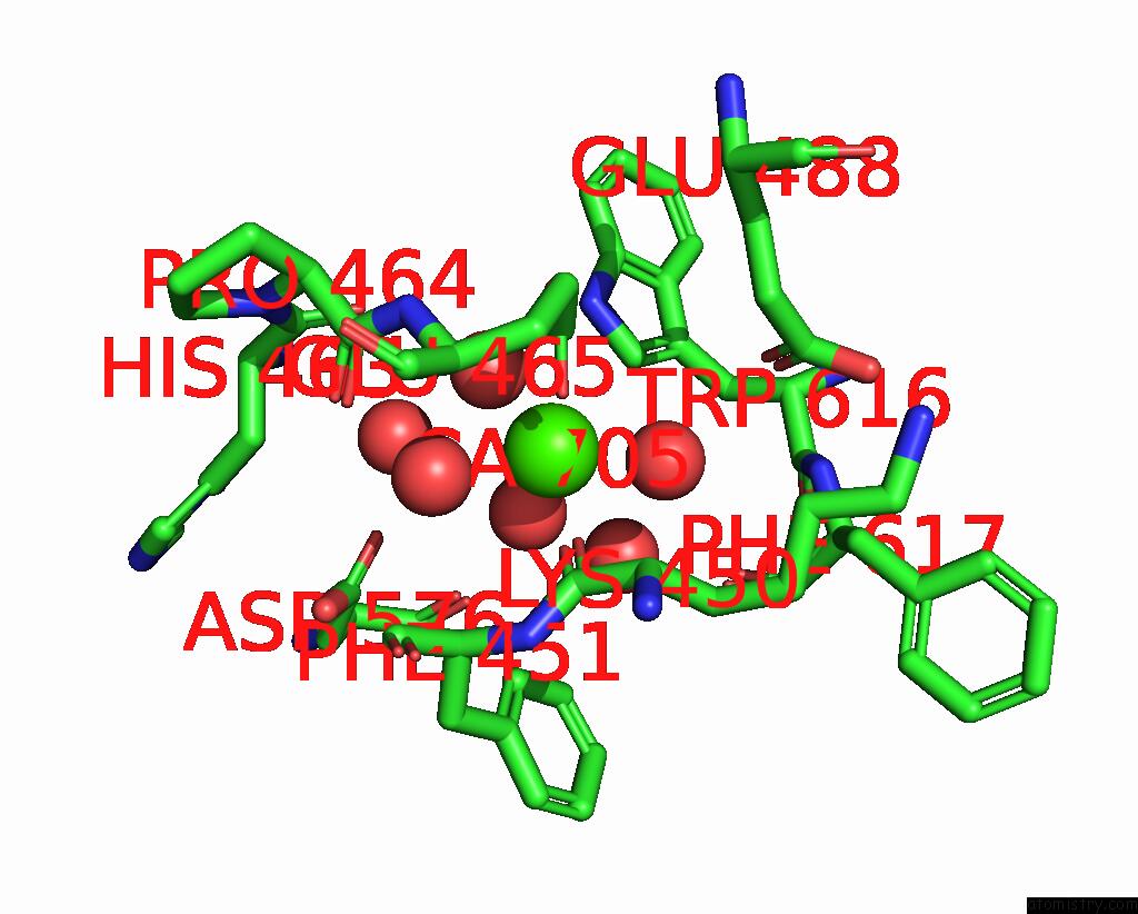
Mono view
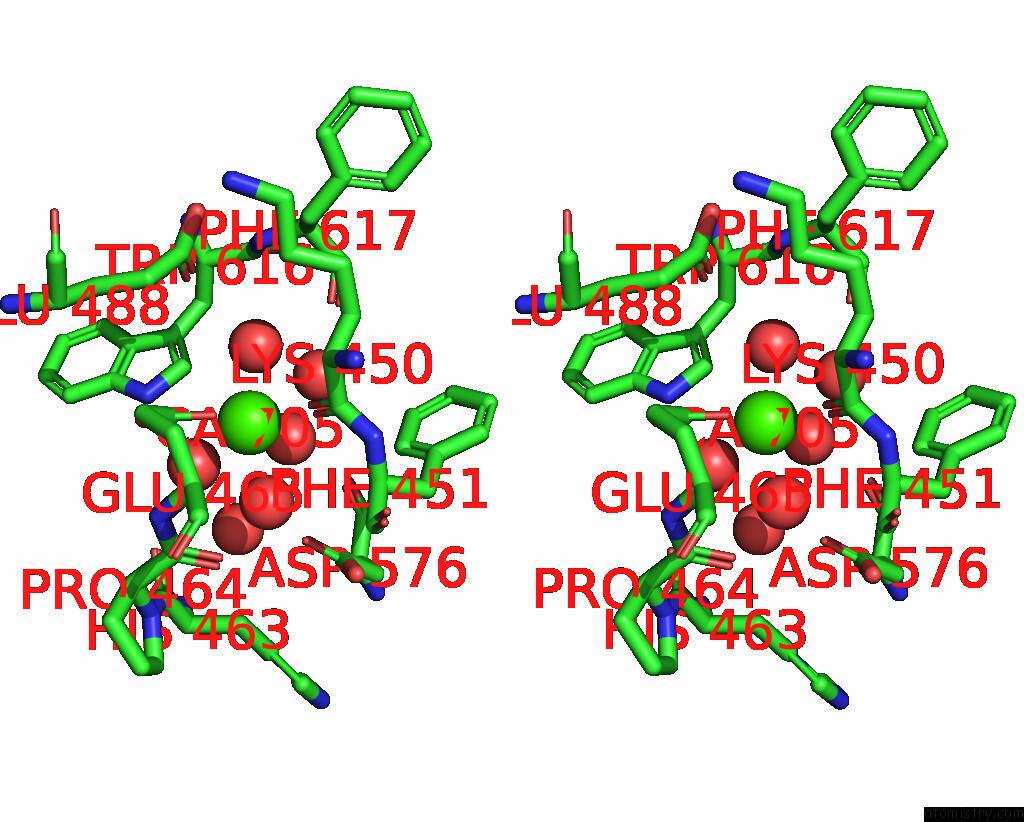
Stereo pair view
|
A full contact list of Calcium with other atoms in the Ca binding
site number 8 of Low-Dose Structure of Marinobacter Nauticus Nitrous Oxide Reductase within 5.0Å range:
probe
|
atom
|
residue
|
distance (Å)
|
B
|
Occ
|
D:Ca705
b:15.3
occ:1.00
|
OE1
|
D:GLU465
|
2.3
|
24.9
|
1.0
|
O
|
D:LYS450
|
2.3
|
15.8
|
1.0
|
O
|
D:HOH1139
|
2.3
|
14.8
|
1.0
|
O
|
C:HOH1165
|
2.3
|
16.1
|
1.0
|
O
|
C:HOH958
|
2.4
|
14.0
|
1.0
|
O
|
D:HOH1242
|
2.5
|
19.2
|
1.0
|
CD
|
D:GLU465
|
3.0
|
23.7
|
1.0
|
OE2
|
D:GLU465
|
3.1
|
32.0
|
1.0
|
C
|
D:LYS450
|
3.4
|
17.0
|
1.0
|
OD1
|
C:ASP576
|
4.1
|
15.9
|
1.0
|
CD1
|
C:TRP616
|
4.2
|
14.7
|
1.0
|
CA
|
D:LYS450
|
4.2
|
14.8
|
1.0
|
O
|
C:HOH997
|
4.4
|
15.6
|
1.0
|
N
|
D:PHE451
|
4.4
|
14.8
|
1.0
|
CG
|
D:GLU465
|
4.5
|
18.9
|
1.0
|
CA
|
D:PHE451
|
4.5
|
15.9
|
1.0
|
NE1
|
C:TRP616
|
4.5
|
14.3
|
1.0
|
CD1
|
D:PHE451
|
4.6
|
19.7
|
1.0
|
O
|
D:HIS463
|
4.6
|
13.8
|
1.0
|
O
|
C:HOH1008
|
4.6
|
16.0
|
1.0
|
OE1
|
D:GLU488
|
4.6
|
19.8
|
1.0
|
O
|
D:PRO464
|
4.7
|
14.2
|
1.0
|
O
|
C:PHE617
|
4.7
|
14.8
|
1.0
|
CB
|
D:LYS450
|
4.7
|
14.8
|
1.0
|
CA
|
D:GLU465
|
5.0
|
14.7
|
1.0
|
|
Reference:
A.Pomowski,
S.Dell'acqua,
A.Wust,
S.R.Pauleta,
I.Moura,
O.Einsle.
Revisiting the Metal Sites of Nitrous Oxide Reductase in A Low-Dose Structure From Marinobacter Nauticus. J.Biol.Inorg.Chem. V. 29 279 2024.
ISSN: ESSN 1432-1327
PubMed: 38720157
DOI: 10.1007/S00775-024-02056-Y
Page generated: Fri Jul 19 13:01:49 2024
|