Calcium »
PDB 9g3k-9ibx »
9g7d »
Calcium in PDB 9g7d: Crystal Structure of Asgpr with Bound Imp
Protein crystallography data
The structure of Crystal Structure of Asgpr with Bound Imp, PDB code: 9g7d
was solved by
H.A.Schreuder,
A.Hofmeister,
with X-Ray Crystallography technique. A brief refinement statistics is given in the table below:
| Resolution Low / High (Å) | 56.85 / 1.59 |
| Space group | C 1 2 1 |
| Cell size a, b, c (Å), α, β, γ (°) | 113.775, 32.689, 40.819, 90, 91.98, 90 |
| R / Rfree (%) | 18.2 / 23.1 |
Calcium Binding Sites:
The binding sites of Calcium atom in the Crystal Structure of Asgpr with Bound Imp
(pdb code 9g7d). This binding sites where shown within
5.0 Angstroms radius around Calcium atom.
In total 3 binding sites of Calcium where determined in the Crystal Structure of Asgpr with Bound Imp, PDB code: 9g7d:
Jump to Calcium binding site number: 1; 2; 3;
In total 3 binding sites of Calcium where determined in the Crystal Structure of Asgpr with Bound Imp, PDB code: 9g7d:
Jump to Calcium binding site number: 1; 2; 3;
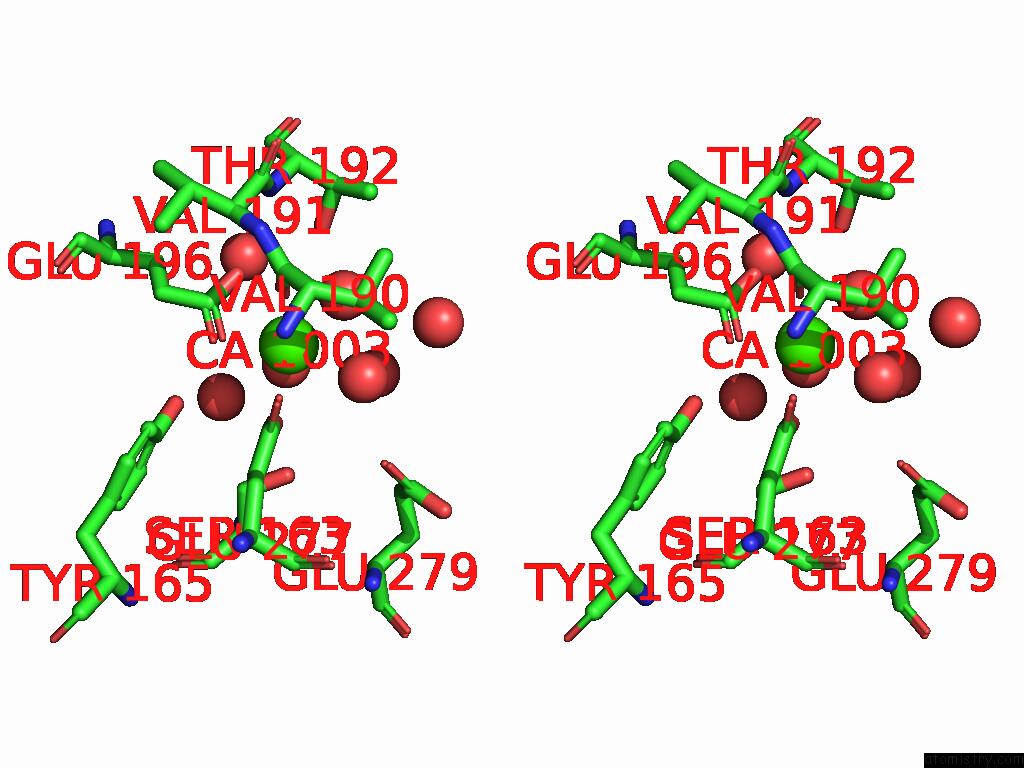
Calcium binding site 1 out of 3 in 9g7d
Go back to
Calcium binding site 1 out
of 3 in the Crystal Structure of Asgpr with Bound Imp
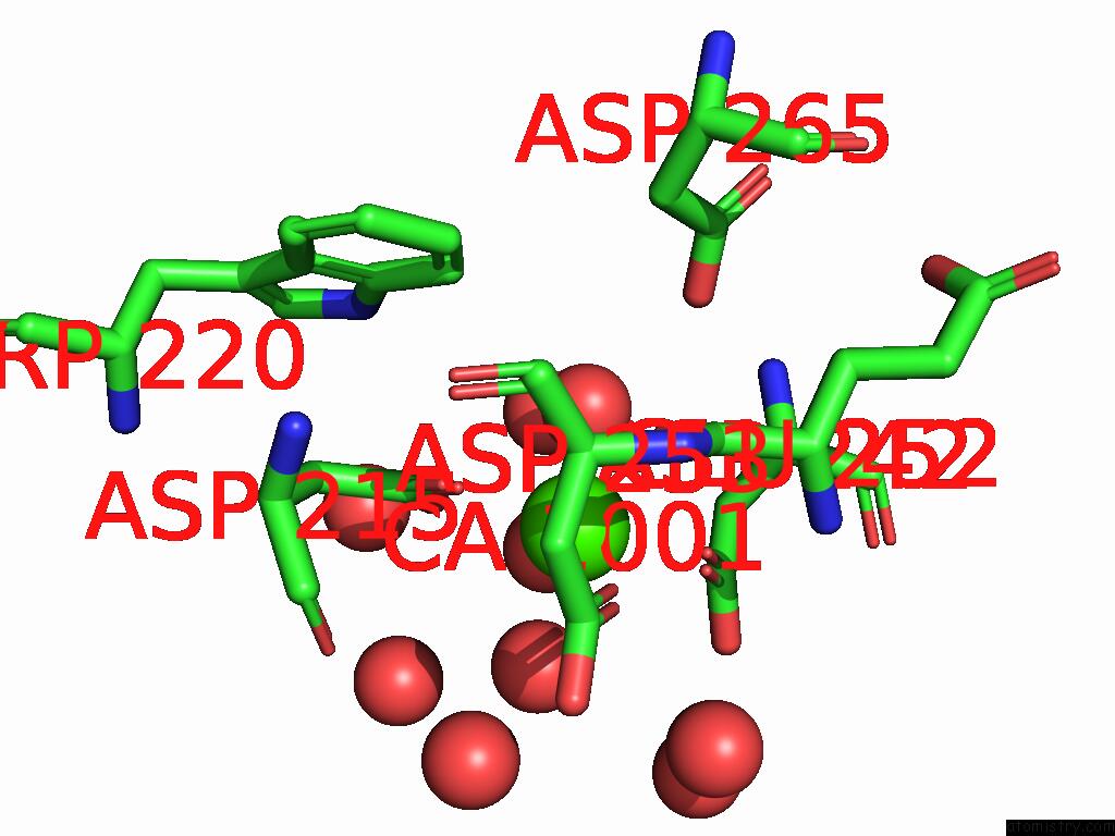
Mono view
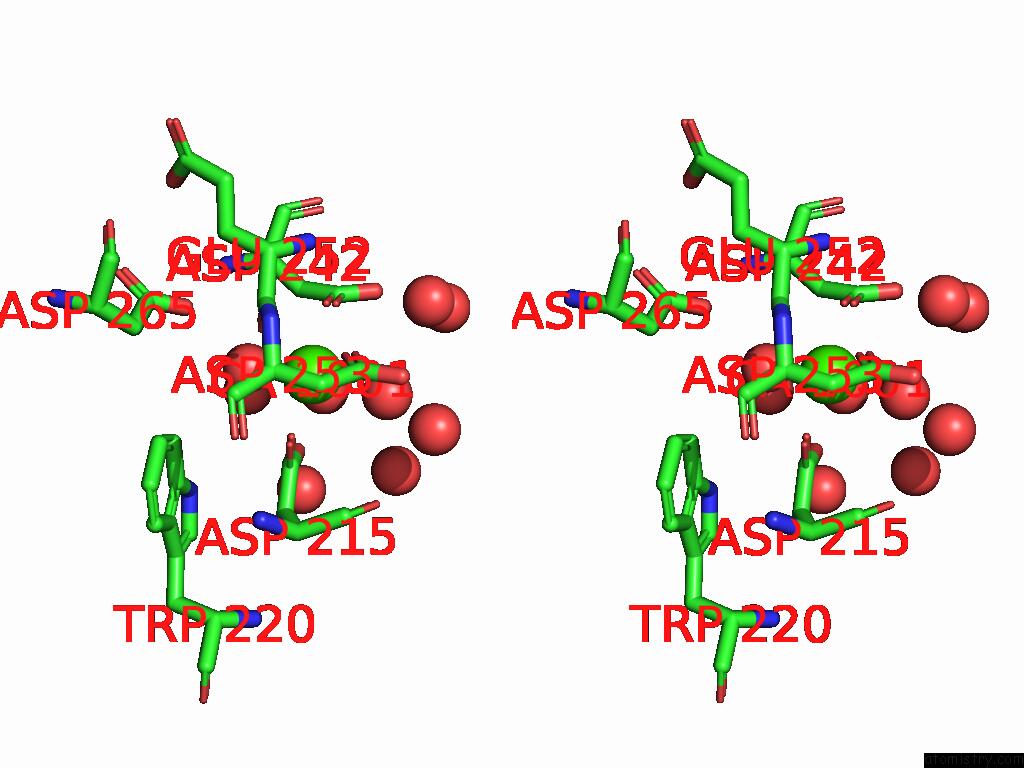
Stereo pair view
Mono view
Stereo pair view
A full contact list of Calcium with other atoms in the Ca binding
site number 1 of Crystal Structure of Asgpr with Bound Imp within 5.0Å range:
|
Calcium binding site 2 out of 3 in 9g7d
Go back to
Calcium binding site 2 out
of 3 in the Crystal Structure of Asgpr with Bound Imp
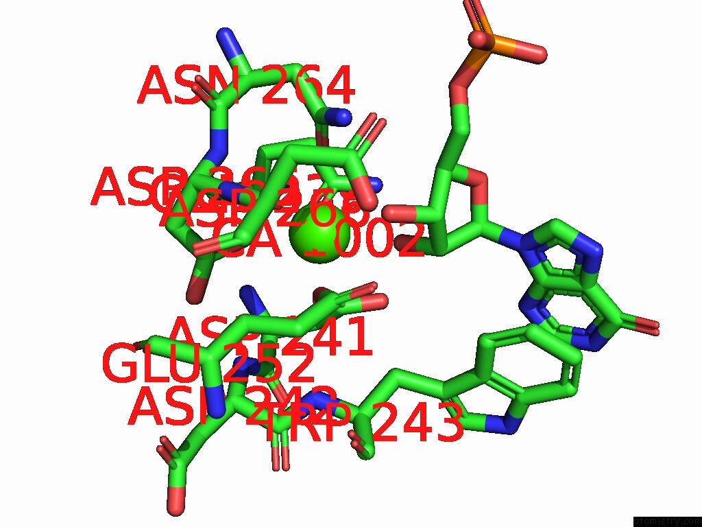
Mono view

Stereo pair view
Mono view
Stereo pair view
A full contact list of Calcium with other atoms in the Ca binding
site number 2 of Crystal Structure of Asgpr with Bound Imp within 5.0Å range:
|
Calcium binding site 3 out of 3 in 9g7d
Go back to
Calcium binding site 3 out
of 3 in the Crystal Structure of Asgpr with Bound Imp
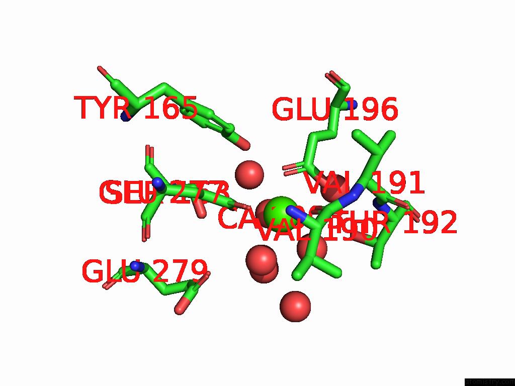
Mono view
Stereo pair view
Mono view
Stereo pair view
A full contact list of Calcium with other atoms in the Ca binding
site number 3 of Crystal Structure of Asgpr with Bound Imp within 5.0Å range:
|
Reference:
A.Hofmeister,
K.Jahn-Hofmann,
B.Brunner,
M.Helms,
C.Metz-Weidmann,
C.Poeverlein,
G.Zech,
Z.Li,
G.Hessler,
H.Schreuder,
B.Elshorst,
A.Krack,
M.Kurz,
C.Heubel,
S.Scheidler.
Trivalent Sirna-Conjugates with Guanosine As Asgpr-Binder Show Potent Knock-Down in Vivo. J.Med.Chem. 2025.
ISSN: ISSN 0022-2623
PubMed: 40052708
DOI: 10.1021/ACS.JMEDCHEM.4C02275
Page generated: Thu Jul 10 09:46:15 2025
ISSN: ISSN 0022-2623
PubMed: 40052708
DOI: 10.1021/ACS.JMEDCHEM.4C02275
Last articles
Cl in 5HGKCl in 5HGI
Cl in 5HG1
Cl in 5HGJ
Cl in 5HDO
Cl in 5HEZ
Cl in 5HG7
Cl in 5HFO
Cl in 5HES
Cl in 5HCJ