Calcium »
PDB 9hh4-9jsk »
9jd0 »
Calcium in PDB 9jd0: Crystal Structure of TMPRSS2 in Complex with Nanobody
Protein crystallography data
The structure of Crystal Structure of TMPRSS2 in Complex with Nanobody, PDB code: 9jd0
was solved by
H.Wang,
Z.Zhao,
X.Liu,
Y.Duan,
H.Yang,
with X-Ray Crystallography technique. A brief refinement statistics is given in the table below:
| Resolution Low / High (Å) | 43.50 / 2.00 |
| Space group | P 41 21 2 |
| Cell size a, b, c (Å), α, β, γ (°) | 137.547, 137.547, 130.325, 90, 90, 90 |
| R / Rfree (%) | 18.8 / 22.1 |
Calcium Binding Sites:
The binding sites of Calcium atom in the Crystal Structure of TMPRSS2 in Complex with Nanobody
(pdb code 9jd0). This binding sites where shown within
5.0 Angstroms radius around Calcium atom.
In total 2 binding sites of Calcium where determined in the Crystal Structure of TMPRSS2 in Complex with Nanobody, PDB code: 9jd0:
Jump to Calcium binding site number: 1; 2;
In total 2 binding sites of Calcium where determined in the Crystal Structure of TMPRSS2 in Complex with Nanobody, PDB code: 9jd0:
Jump to Calcium binding site number: 1; 2;
Calcium binding site 1 out of 2 in 9jd0
Go back to
Calcium binding site 1 out
of 2 in the Crystal Structure of TMPRSS2 in Complex with Nanobody
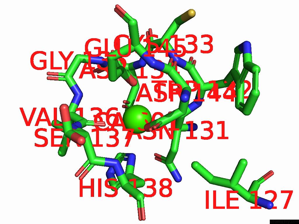
Mono view
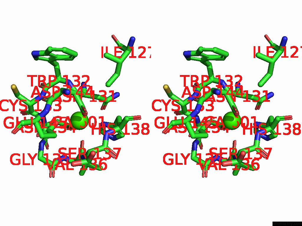
Stereo pair view
Mono view
Stereo pair view
A full contact list of Calcium with other atoms in the Ca binding
site number 1 of Crystal Structure of TMPRSS2 in Complex with Nanobody within 5.0Å range:
|
Calcium binding site 2 out of 2 in 9jd0
Go back to
Calcium binding site 2 out
of 2 in the Crystal Structure of TMPRSS2 in Complex with Nanobody
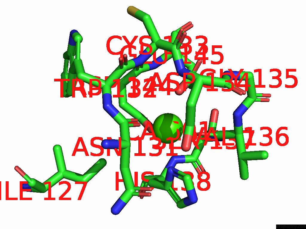
Mono view
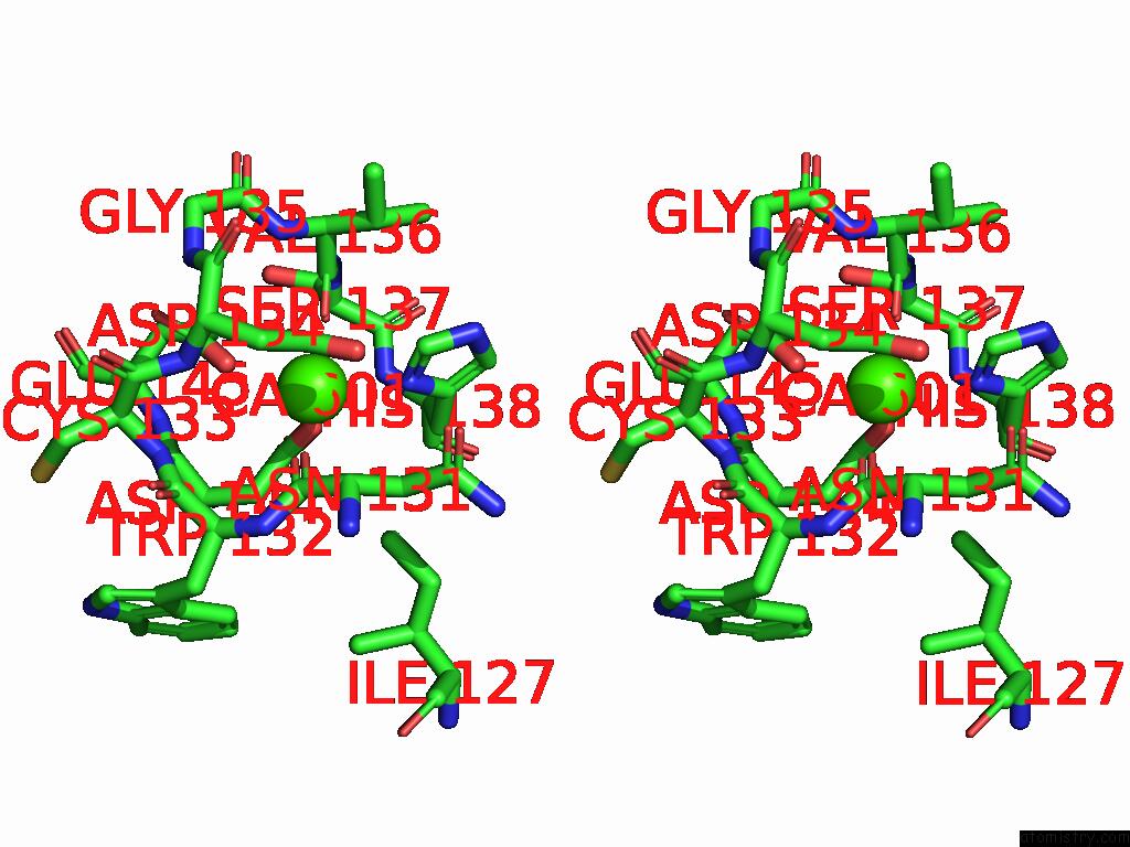
Stereo pair view
Mono view
Stereo pair view
A full contact list of Calcium with other atoms in the Ca binding
site number 2 of Crystal Structure of TMPRSS2 in Complex with Nanobody within 5.0Å range:
|
Reference:
Z.Zhao,
Q.Yang,
X.Liu,
M.Li,
Y.Duan,
M.Du,
A.Zhou,
H.Liu,
Y.He,
W.Wang,
Y.Lu,
X.Zhang,
H.Wang,
X.Yang,
H.Zhang,
X.Chen,
Z.Rao,
H.Yang.
The Crystal Structure of Coronavirus Rbd-TMPRSS2 Complex Provides Basis For the Discovery of Therapeutic Antibodies. Nat Commun V. 16 6636 2025.
ISSN: ESSN 2041-1723
PubMed: 40681508
DOI: 10.1038/S41467-025-62023-2
Page generated: Fri Aug 22 22:37:15 2025
ISSN: ESSN 2041-1723
PubMed: 40681508
DOI: 10.1038/S41467-025-62023-2
Last articles
Mn in 9LJUMn in 9LJW
Mn in 9LJS
Mn in 9LJR
Mn in 9LJT
Mn in 9LJV
Mg in 9UA2
Mg in 9R96
Mg in 9VM1
Mg in 9P01