Calcium »
PDB 1aui-1b82 »
1aux »
Calcium in PDB 1aux: Structure of the C Domain of Synapsin Ia From Bovine Brain with Calcium Atp-Gamma-S Bound
Protein crystallography data
The structure of Structure of the C Domain of Synapsin Ia From Bovine Brain with Calcium Atp-Gamma-S Bound, PDB code: 1aux
was solved by
L.Esser,
C.Wang,
J.Deisenhofer,
with X-Ray Crystallography technique. A brief refinement statistics is given in the table below:
| Resolution Low / High (Å) | 20.00 / 2.30 |
| Space group | P 32 2 1 |
| Cell size a, b, c (Å), α, β, γ (°) | 76.160, 76.160, 182.330, 90.00, 90.00, 120.00 |
| R / Rfree (%) | 20.6 / 26.9 |
Calcium Binding Sites:
The binding sites of Calcium atom in the Structure of the C Domain of Synapsin Ia From Bovine Brain with Calcium Atp-Gamma-S Bound
(pdb code 1aux). This binding sites where shown within
5.0 Angstroms radius around Calcium atom.
In total 2 binding sites of Calcium where determined in the Structure of the C Domain of Synapsin Ia From Bovine Brain with Calcium Atp-Gamma-S Bound, PDB code: 1aux:
Jump to Calcium binding site number: 1; 2;
In total 2 binding sites of Calcium where determined in the Structure of the C Domain of Synapsin Ia From Bovine Brain with Calcium Atp-Gamma-S Bound, PDB code: 1aux:
Jump to Calcium binding site number: 1; 2;
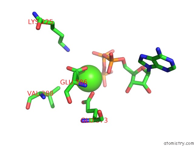
Calcium binding site 1 out of 2 in 1aux
Go back to
Calcium binding site 1 out
of 2 in the Structure of the C Domain of Synapsin Ia From Bovine Brain with Calcium Atp-Gamma-S Bound

Mono view

Stereo pair view
Mono view
Stereo pair view
A full contact list of Calcium with other atoms in the Ca binding
site number 1 of Structure of the C Domain of Synapsin Ia From Bovine Brain with Calcium Atp-Gamma-S Bound within 5.0Å range:
|
Calcium binding site 2 out of 2 in 1aux
Go back to
Calcium binding site 2 out
of 2 in the Structure of the C Domain of Synapsin Ia From Bovine Brain with Calcium Atp-Gamma-S Bound
Mono view

Stereo pair view
Mono view
Stereo pair view
A full contact list of Calcium with other atoms in the Ca binding
site number 2 of Structure of the C Domain of Synapsin Ia From Bovine Brain with Calcium Atp-Gamma-S Bound within 5.0Å range:
|
Reference:
L.Esser,
C.R.Wang,
M.Hosaka,
C.S.Smagula,
T.C.Sudhof,
J.Deisenhofer.
Synapsin I Is Structurally Similar to Atp-Utilizing Enzymes. Embo J. V. 17 977 1998.
ISSN: ISSN 0261-4189
PubMed: 9463376
DOI: 10.1093/EMBOJ/17.4.977
Page generated: Mon Jul 7 13:28:11 2025
ISSN: ISSN 0261-4189
PubMed: 9463376
DOI: 10.1093/EMBOJ/17.4.977
Last articles
Mg in 6T2CMg in 6T1Y
Mg in 6T23
Mg in 6T20
Mg in 6T0V
Mg in 6T0S
Mg in 6T15
Mg in 6T0N
Mg in 6T0K
Mg in 6T0J